
| 应用 | 稀释度 |
|---|---|
| IHC-P | 1:3200 |
| WB | 1:200 |
| IP | 1:25 |

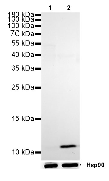
WB result of Calprotectin Rabbit mAb
Primary antibody: Calprotectin Rabbit mAb at 1/200 dilution
Lane 1: C2C12 whole cell lysate 20 µg
Lane 2: THP-1 whole cell lysate 20 µg
Negative control: C2C12 whole cell lysate
Secondary antibody: Goat Anti-Rabbit IgG, (H+L), HRP conjugated at 1/10000 dilution
Predicted MW: 11 kDa
Observed MW: 11 kDa
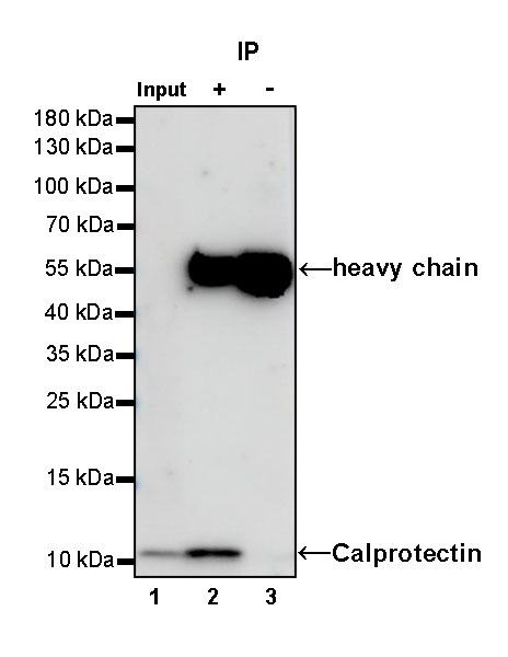
Calprotectin Rabbit mAb at 1/25 dilution (0.4 µg) immunoprecipitating Calprotectin in 0.4 mg THP-1 whole cell lysate.
Western blot was performed on the immunoprecipitate using Calprotectin Rabbit mAb at 1/1000 dilution.
Secondary antibody (HRP) for IP was used at 1/400 dilution.
Lane 1: THP-1 whole cell lysate 5 µg (Input)
Lane 2: Calprotectin Rabbit mAb IP in THP-1 whole cell lysate
Lane 3: Rabbit monoclonal IgG IP in THP-1 whole cell lysate
Predicted MW: 11 kDa
Observed MW: 11 kDa
(This blot was developed with high sensitivity substrate)
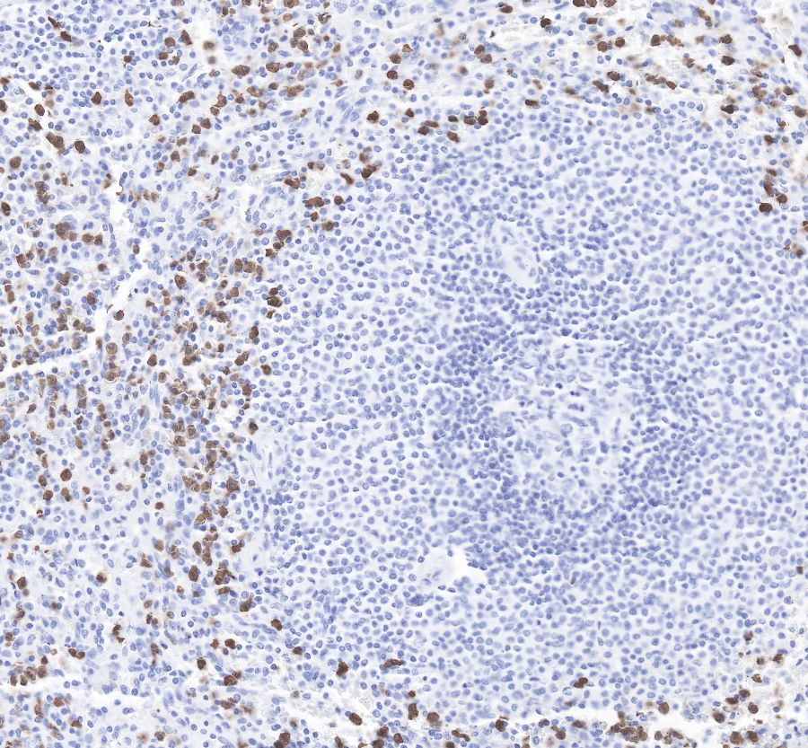
IHC shows positive staining in paraffin-embedded human spleen. Anti-Synaptophysin antibody was used at 1/3200 dilution, followed by a Goat Anti-Rabbit IgG H&L (HRP) ready to use.
Counterstained with hematoxylin.
Heat mediated antigen retrieval with Tris/EDTA buffer pH9.0 was performed before commencing with IHC staining protocol.
IHC shows positive staining in paraffin-embedded human stomach. Anti-Synaptophysin antibody was used at 1/3200 dilution, followed by a Goat Anti-Rabbit IgG H&L (HRP) ready to use.
Counterstained with hematoxylin.
Heat mediated antigen retrieval with Tris/EDTA buffer pH9.0 was performed before commencing with IHC staining protocol.
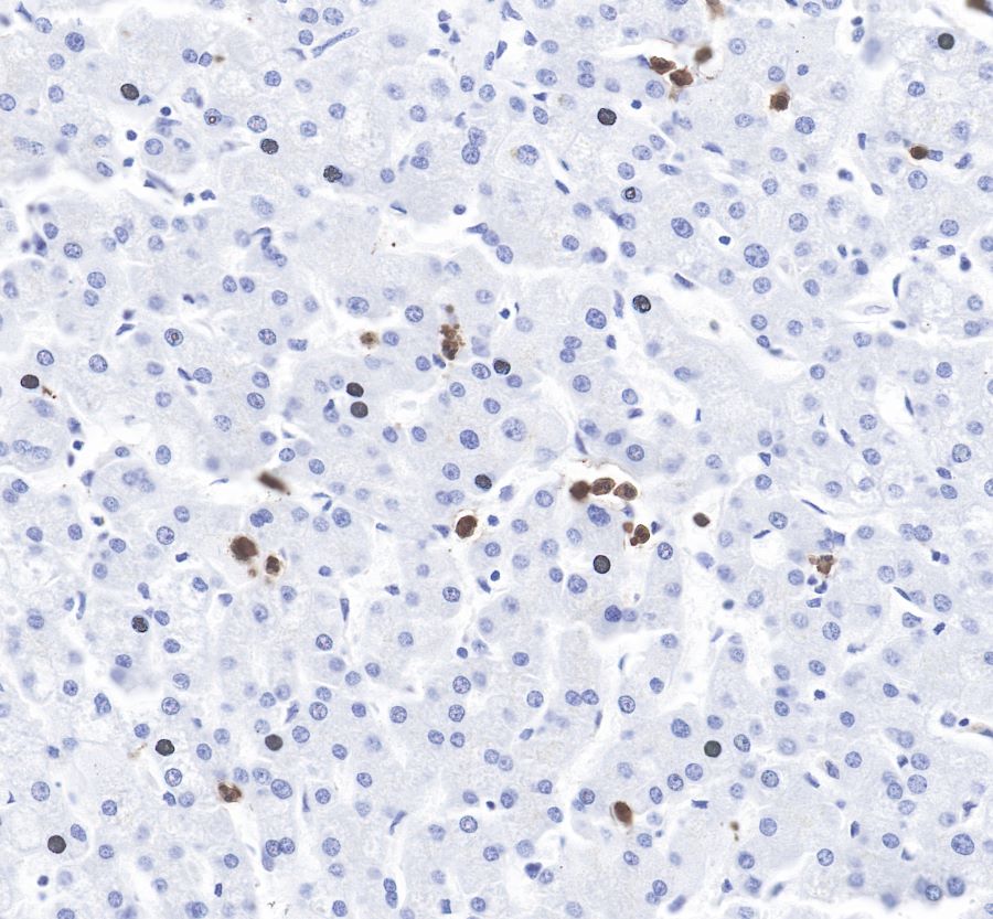
IHC shows positive staining in paraffin-embedded human liver.
Anti-Synaptophysin antibody was used at 1/3200 dilution, followed by a Goat Anti-Rabbit IgG H&L (HRP) ready to use.
Counterstained with hematoxylin.
Heat mediated antigen retrieval with Tris/EDTA buffer pH9.0 was performed before commencing with IHC staining protocol.
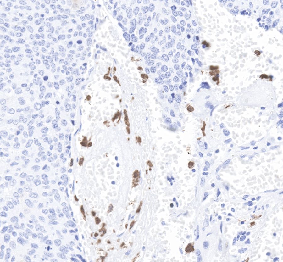
IHC shows positive staining in paraffin-embedded human cervix cancer.
Anti-Synaptophysin antibody was used at 1/3200 dilution, followed by a Goat Anti-Rabbit IgG H&L (HRP) ready to use.
Counterstained with hematoxylin.
Heat mediated antigen retrieval with Tris/EDTA buffer pH9.0 was performed before commencing with IHC staining protocol.
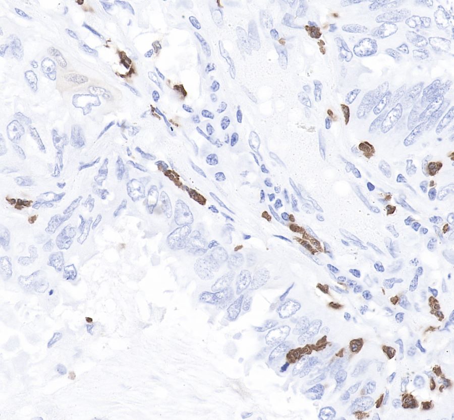
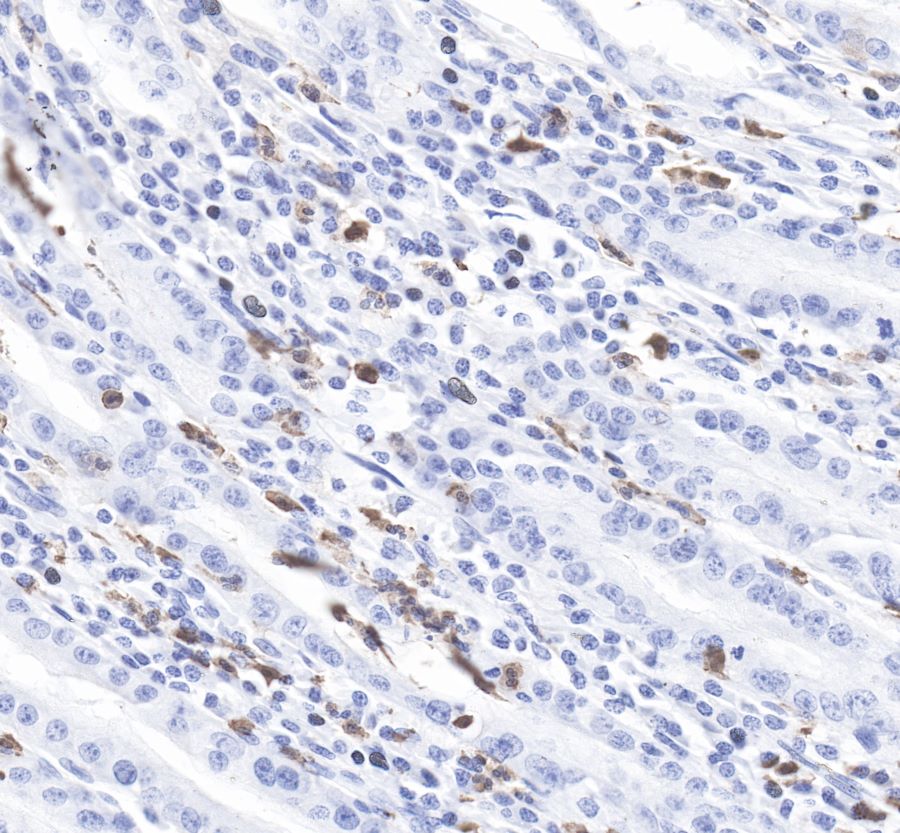
IHC shows positive staining in paraffin-embedded human lung cancer.
Anti-Synaptophysin antibody was used at 1/3200 dilution, followed by a Goat Anti-Rabbit IgG H&L (HRP) ready to use.
Counterstained with hematoxylin.
Heat mediated antigen retrieval with Tris/EDTA buffer pH9.0 was performed before commencing with IHC staining protocol.
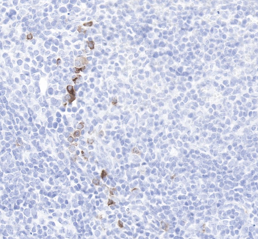
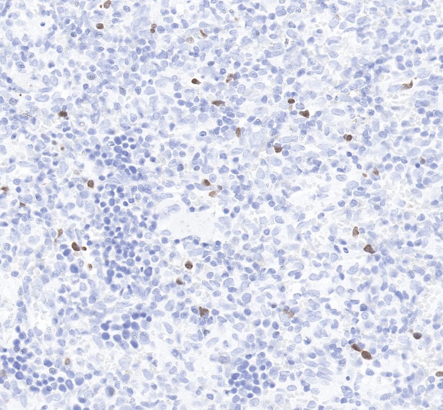
IHC shows positive staining in paraffin-embedded mouse spleen. Anti-Synaptophysin antibody was used at 1/3200 dilution, followed by a Goat Anti-Rabbit IgG H&L (HRP) ready to use.
Counterstained with hematoxylin.
Heat mediated antigen retrieval with Tris/EDTA buffer pH9.0 was performed before commencing with IHC staining protocol.
IHC shows positive staining in paraffin-embedded rat spleen.
Anti-Synaptophysin antibody was used at 1/3200 dilution, followed by a Goat Anti-Rabbit IgG H&L (HRP) ready to use.
Counterstained with hematoxylin. Heat mediated antigen retrieval with Tris/EDTA buffer pH9.0 was performed before commencing with IHC staining protocol.
 您现在的位置: