

| 应用 | 稀释度 |
|---|---|
| IHC-P | 1:1000 |
| WB | 1:500 |
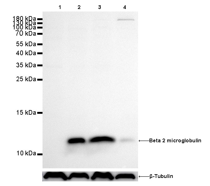
WB result of beta 2 microglobulin Rabbit mAb Primary antibody: beta 2 microglobulin Rabbit mAb at 1/500 dilution
Lane 1: Daudi whole cell lysate 20 µg
Lane 2: Hela whole cell lysate 20 µg
Lane 3: Jurkat whole cell lysate 20 µg
Lane 4: HepG2 whole cell lysate 20 µg
Negative control: Daudi whole cell lysate Secondary antibody: Goat Anti-Rabbit IgG, (H+L), HRP conjugated at 1/10000 dilution
Predicted MW: 12 kDa
Observed MW: 12 kDa
Exposure time: 180s
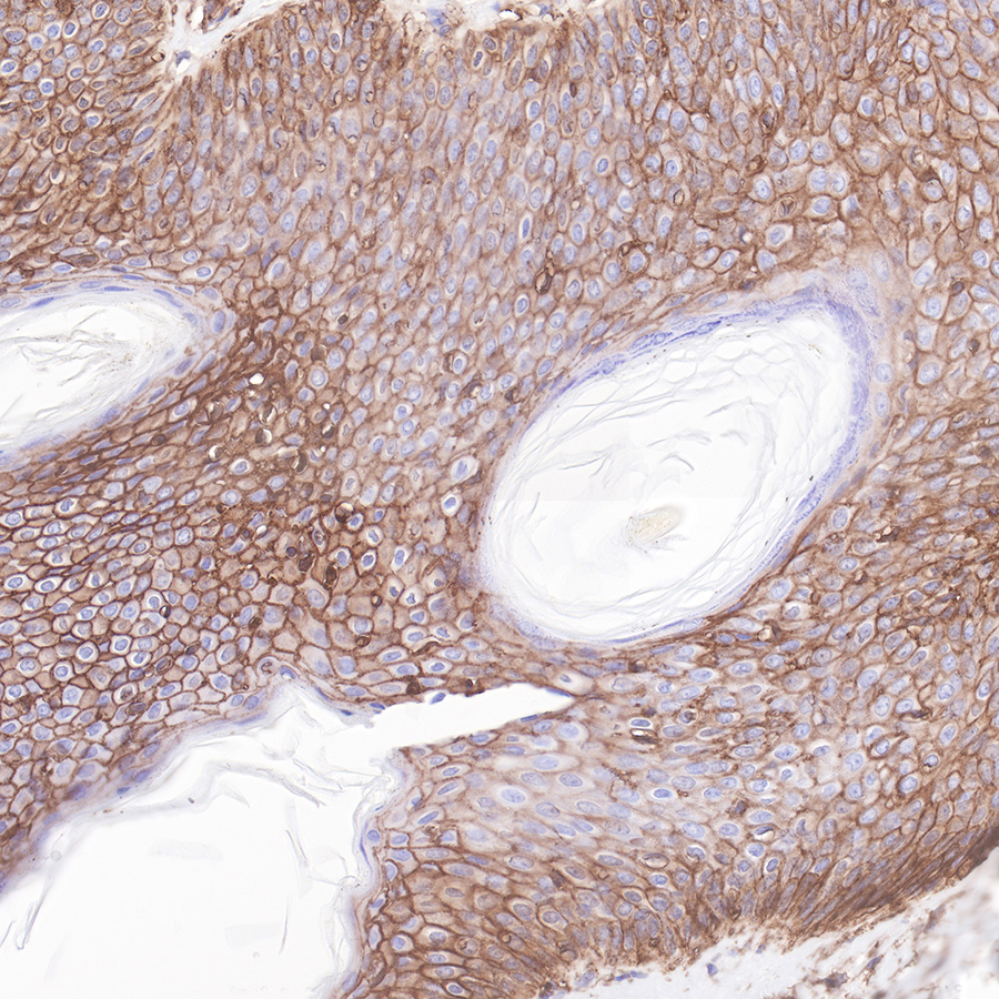
IHC shows positive staining in paraffin-embedded human skin.
Anti-Beta 2 microglobulin antibody was used at 1/1000 dilution, followed by a Goat Anti-Rabbit IgG H&L (HRP) ready to use.
Counterstained with hematoxylin.
Heat mediated antigen retrieval with Tris/EDTA buffer pH9.0 was performed before commencing with IHC staining protocol.
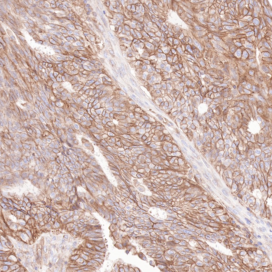
IHC shows positive staining in paraffin-embedded human ovarian cancer.
Anti-Beta 2 microglobulin antibody was used at 1/1000 dilution, followed by a Goat Anti-Rabbit IgG H&L (HRP) ready to use. Counterstained with hematoxylin.
Heat mediated antigen retrieval with Tris/EDTA buffer pH9.0 was performed before commencing with IHC staining protocol.
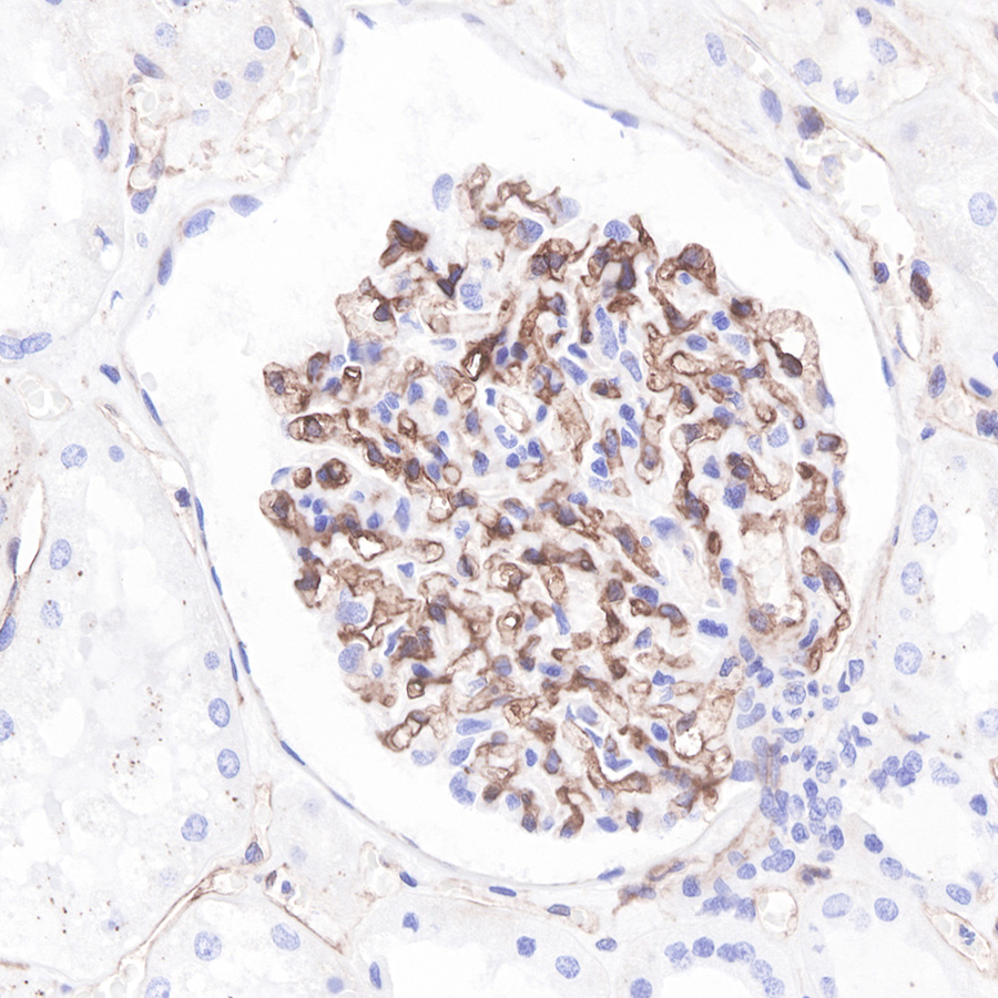
IHC shows positive staining in paraffin-embedded human kidney. Anti-Beta 2 microglobulin antibody was used at 1/1000 dilution, followed by a Goat Anti-Rabbit IgG H&L (HRP) ready to use.
Counterstained with hematoxylin.
Heat mediated antigen retrieval with Tris/EDTA buffer pH9.0 was performed before commencing with IHC staining protocol.
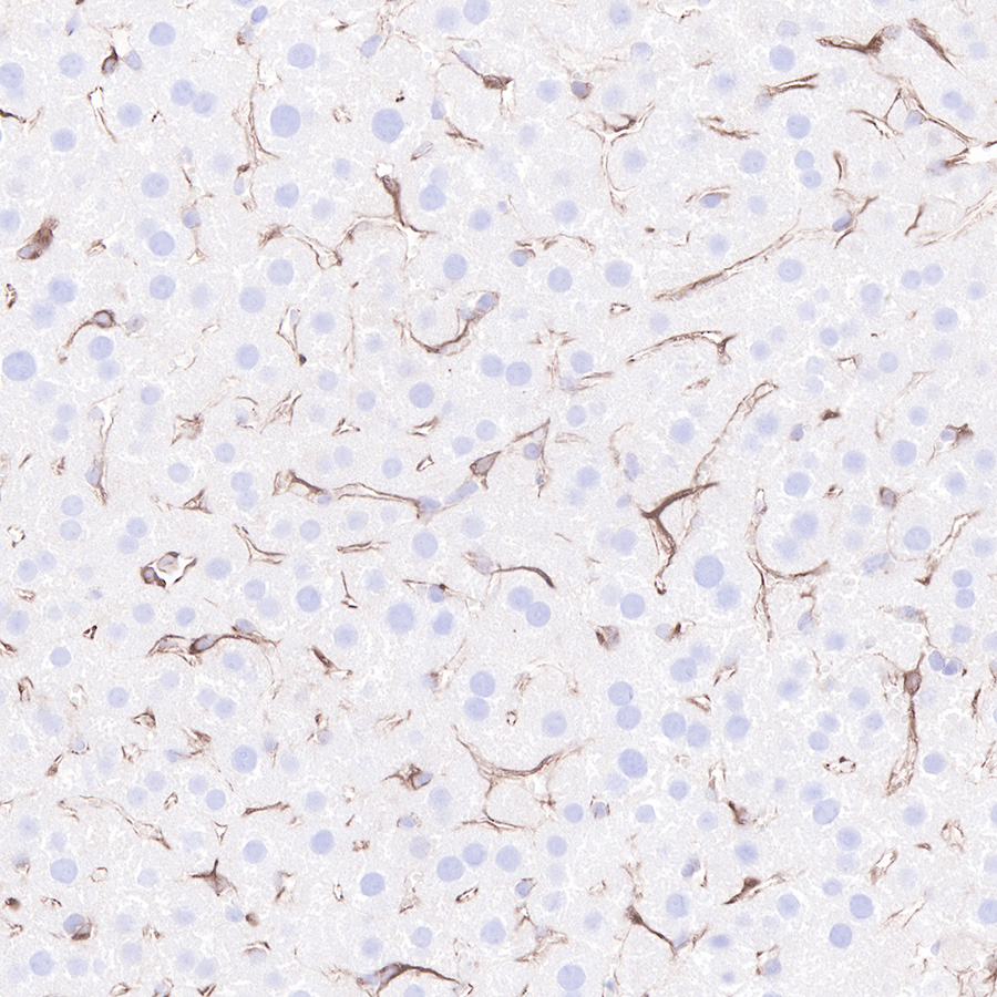
IHC shows positive staining in paraffin-embedded mouse liver.
Anti-Beta 2 microglobulin antibody was used at 1/1000 dilution, followed by a Goat Anti-Rabbit IgG H&L (HRP) ready to use.
Counterstained with hematoxylin.
Heat mediated antigen retrieval with Tris/EDTA buffer pH9.0 was performed before commencing with IHC staining protocol.
 您现在的位置: