

12 months from date of receipt / reconstitution, -20 °C as supplied
| 应用 | 稀释度 |
|---|---|
| IHC-P | 1:1000 |
| WB | 1:500 |
| ICC | 1:250 |
Desmin is a 53 kDa muscle -specific molecule, which belongs to the middle silk super family of cell skeleton protein. Cyclical protein digosum formation forms a stable wire -shaped network, which can stabilize the horizontal arrangement of the muscle fiber. Most mutations affect filament assembly. Desmin knockout mice will form myocardial disease, skeletal muscle defects and smooth muscle defects. It is a good screening symbol of tumor with muscle -derived differentiation, including transverse muscle sarcoma, horizontal fibroids, smooth muscle sarcoma, smooth fibroids, smooth muscles, and horizontal muscle cells in other tumors.
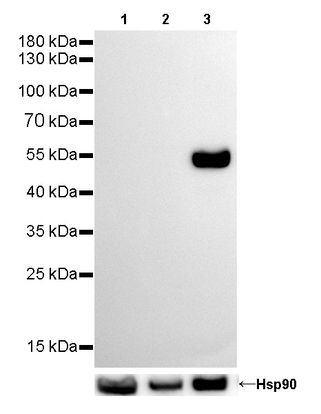
WB result of Desmin Rabbit mAb
Primary antibody: Desmin Rabbit mAb at 1/500 dilution
Lane 1: Hela whole cell lysate 20 µg
Lane 2: A431 whole cell lysate 20 µg
Lane 3: C2C12 whole cell lysate 20 µg
Negative control: Hela whole cell lysate
Low expression control: A431 whole cell lysate
Secondary antibody: Goat Anti-Rabbit IgG, (H+L), HRP conjugated at 1/10000 dilution
Predicted MW: 53 kDa
Observed MW: 53 kDa
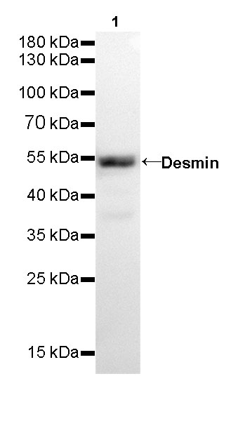
WB result of Desmin Rabbit mAb
Primary antibody: Desmin Rabbit mAb at 1/500 dilution
Lane 1: mouse heart lysate 20 µg
Secondary antibody: Goat Anti-Rabbit IgG, (H+L), HRP conjugated at 1/10000 dilution
Predicted MW: 53 kDa
Observed MW: 53 kDa
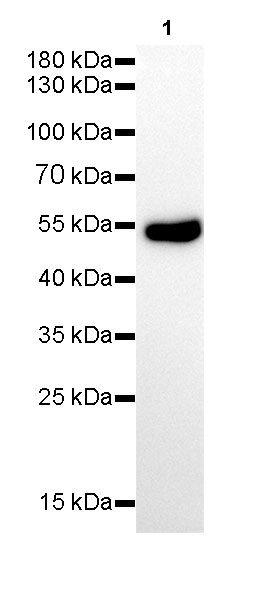
WB result of Desmin Rabbit mAb
Primary antibody: Desmin Rabbit mAb at 1/500 dilution
Lane 1: rat heart whole cell lysate 20 µg
Secondary antbody: Goat Anti-Rabbit IgG, (H+L), HRP conjugated at 1/10000 dilution
Predicted MW: 53 kDa
Observed MW: 53 kDa
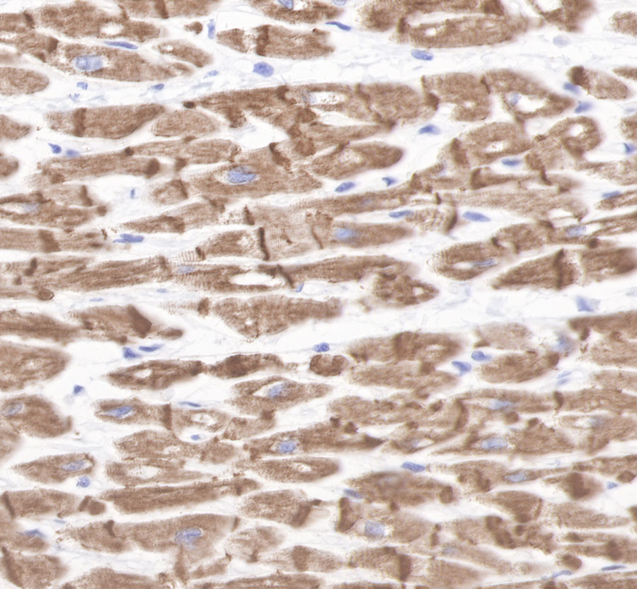
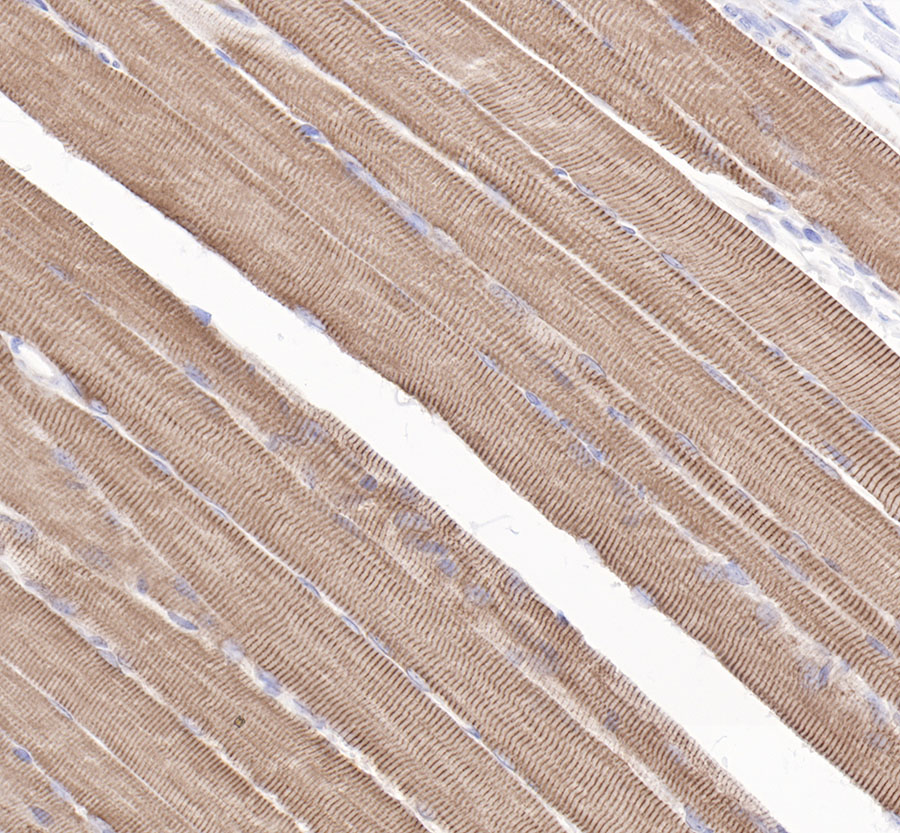
IHC shows positive staining in paraffin-embedded human heart.
Anti-Desmin antibody was used at 1/1000 dilution, followed by a Goat Anti-Rabbit IgG H&L (HRP) ready to use.
Counterstained with hematoxylin.
Heat mediated antigen retrieval with Tris/EDTA buffer pH9.0 was performed before commencing with IHC staining protocol.
IHC shows positive staining in paraffin-embedded human skeletal muscle.
Anti-Desmin antibody was used at 1/1000 dilution, followed by a Goat Anti-Rabbit IgG H&L (HRP) ready to use.
Counterstained with hematoxylin.
Heat mediated antigen retrieval with Tris/EDTA buffer pH9.0 was performed before commencing with IHC staining protocol.
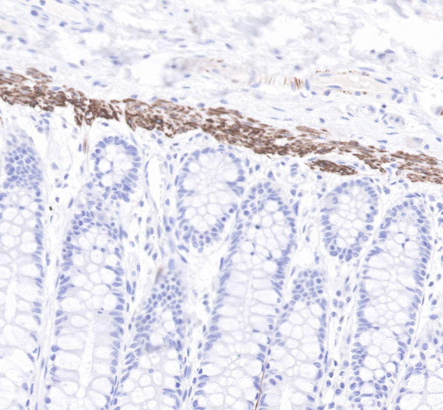
IHC shows positive staining in paraffin-embedded human colon.
Anti-Desmin antibody was used at 1/1000 dilution, followed by a Goat Anti-Rabbit IgG H&L (HRP) ready to use.
Counterstained with hematoxylin.
Heat mediated antigen retrieval with Tris/EDTA buffer pH9.0 was performed before commencing with IHC staining protocol.
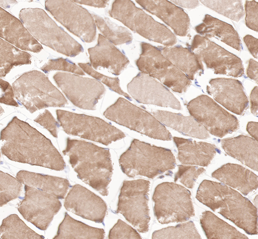
IHC shows positive staining in paraffin-embedded mouse skeletal muscle.
Anti-Desmin antibody was used at 1/1000 dilution, followed by a Goat Anti-Rabbit IgG H&L (HRP) ready to use.
Counterstained with hematoxylin.
Heat mediated antigen retrieval with Tris/EDTA buffer pH9.0 was performed before commencing with IHC staining protocol.
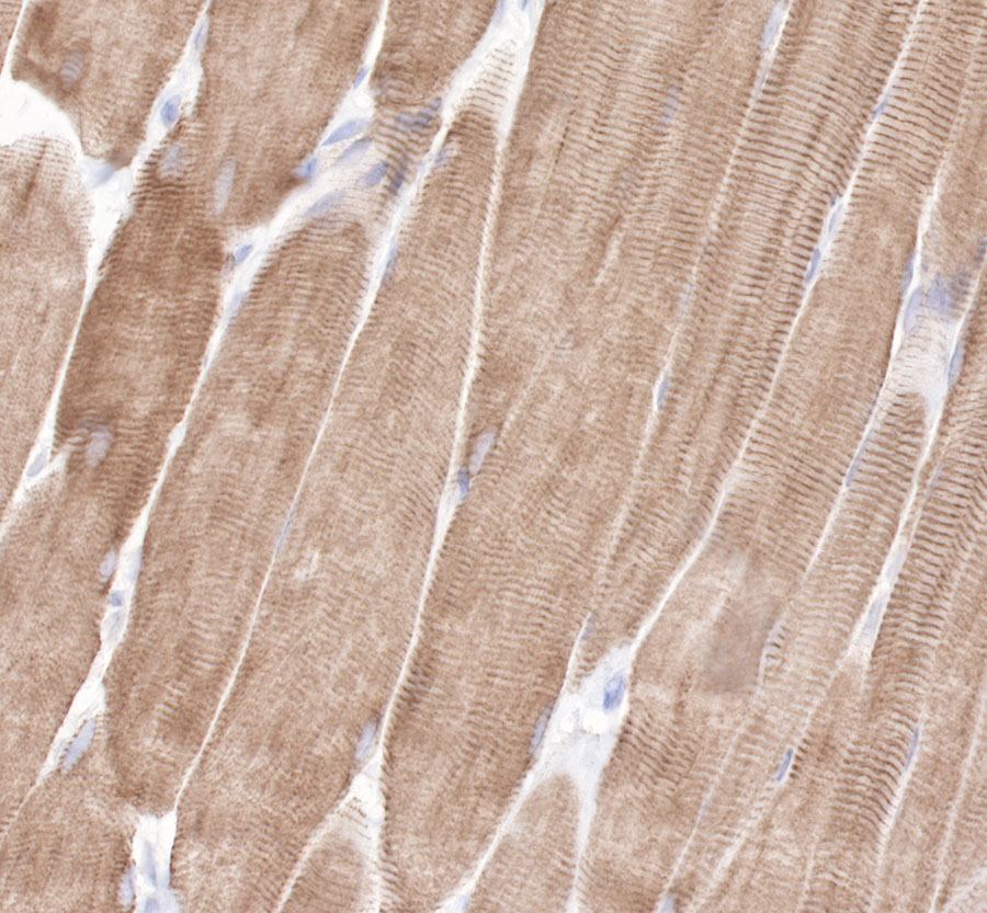
IHC shows positive staining in paraffin-embedded rat skeletal muscle.
Anti-Desmin antibody was used at 1/1000 dilution, followed by a Goat Anti-Rabbit IgG H&L (HRP) ready to use.
Counterstained with hematoxylin.
Heat mediated antigen retrieval with Tris/EDTA buffer pH9.0 was performed before commencing with IHC staining protocol.
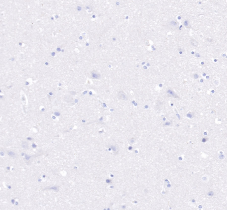
IHC shows negative staining in paraffin-embedded human brain.
Anti-Desmin antibody was used at 1/1000 dilution, followed by a Goat Anti-Rabbit IgG H&L (HRP) ready to use.
Counterstained with hematoxylin.
Heat mediated antigen retrieval with Tris/EDTA buffer pH9.0 was performed before commencing with IHC staining protocol.
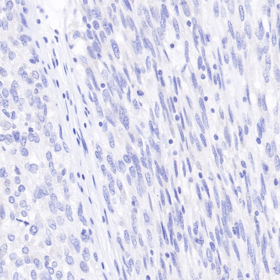
Negative control.IHC shows negative staining in paraffin-embedded human GIST.
Anti-Desmin antibody was used at 1/1000 dilution, followed by a Goat Anti-Rabbit IgG H&L (HRP) ready to use.
Counterstained with hematoxylin.
Heat mediated antigen retrieval with Tris/EDTA buffer pH9.0 was performed before commencing with IHC staining protocol.
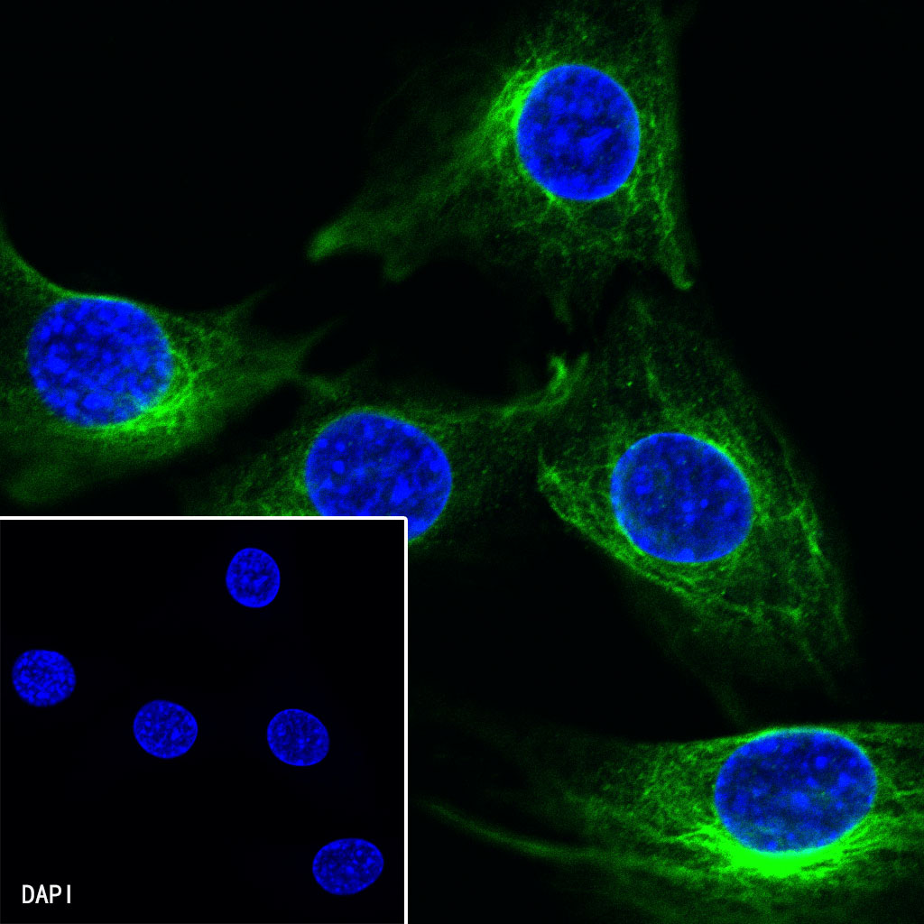
ICC shows positive staining in C2C12 cells. Anti-Desmin antibody was used at 1/250 dilution (Green) and incubated overnight at 4°C. Goat polyclonal Antibody to Rabbit IgG - H&L (Alexa Fluor® 488) was used as secondary antibody at 1/1000 dilution. The cells were fixed with 4%PFA and permeabilized with 0.1% PBS-Triton X-100. Nuclei were counterstained with DAPI (Blue).
 您现在的位置: