
| 应用 | 稀释度 |
|---|---|
| IHC-P | 1:2000 |
| WB | 1:1000 |
| IP | 1:25 |
| FACS | 1:500 |
| ICC | 1:500 |
CD14 (cluster of differentiation 14) is a human protein made mostly by macrophages as part of the innate immune system. It helps to detect bacteria in the body by binding lipopolysaccharide (LPS), a pathogen-associated molecular pattern (PAMP). CD14 exists in two forms, one anchored to the membrane by a glycosylphosphatidylinositol (GPI) tail (mCD14), the other a soluble form (sCD14). Soluble CD14 either appears after shedding of mCD14 (48 kDa) or is directly secreted from intracellular vesicles (56 kDa).

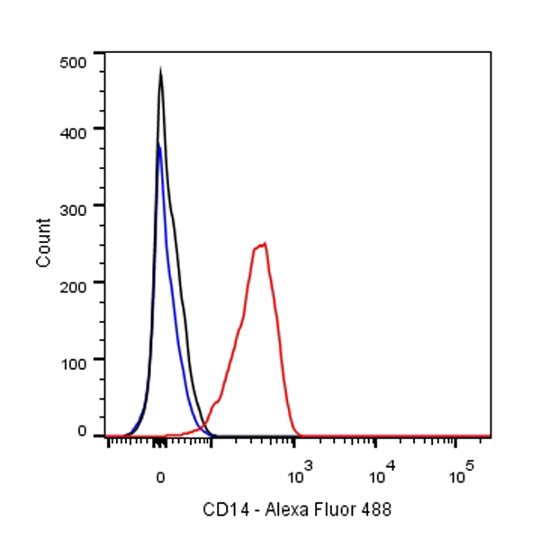
Flow cytometric analysis of THP-1 cells labelled with CD14 antibody at 1/50 dilution (1 μg)/ (red) compared with a Rabbit monoclonal IgG (Black) isotype control and an unlabelled control (cells without incubation with primary antibody and secondary antibody) (Blue).
Goat Anti-Rabbit IgG Alexa Fluor® 488 at 1/1000 dilution was used as the secondary antibody

WB result of CD14 Rabbit mAb
Primary antibody: CD14 Rabbit mAb at 1/1000 dilution
Lane 1: Neuro-2a whole cell lysate 20 µg
Lane 2: THP-1 whole cell lysate 20 µg
Lane 3: A549 whole cell lysate 20 µg
Lane 4: HL-60 whole cell lysate 20 µg
Negative control: Neuro-2a whole cell lysate
Secondary antibody: Goat Anti-Rabbit IgG, (H+L), HRP conjugated at 1/10000 dilution
Predicted MW: 40 kDa
Observed MW: 47~57 kDa
Exposure time: Lane 1、lane 2 and lane 3: 120s; Lane 4: 30s
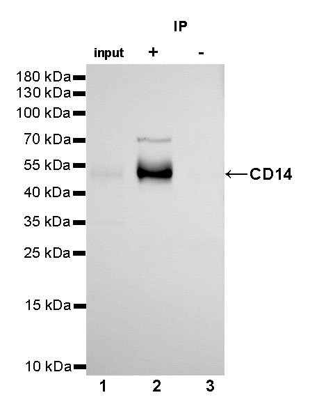
CD14 Rabbit mAb at 1/25 dilution (2µg) immunoprecipitating CD14 in 0.4mg A549 whole cell lysate.
Western blot was performed on the immunoprecipitate using CD14 Rabbit mAb at 1/1000 dilution.
Secondary antibody (HRP) for IP was used at 1/400 dilution.
Lane 1 : A549 whole cell lysate 10µg (input)
Lane 2 : CD14 Rabbit mAb IP in A549 whole cell lysate
Lane 3 : Rabbit monoclonal IgG IP in A549 whole cell lysate
Predicted MW: 40 kDa
Observed MW: 47~57 kDa
Exposure time: 180s
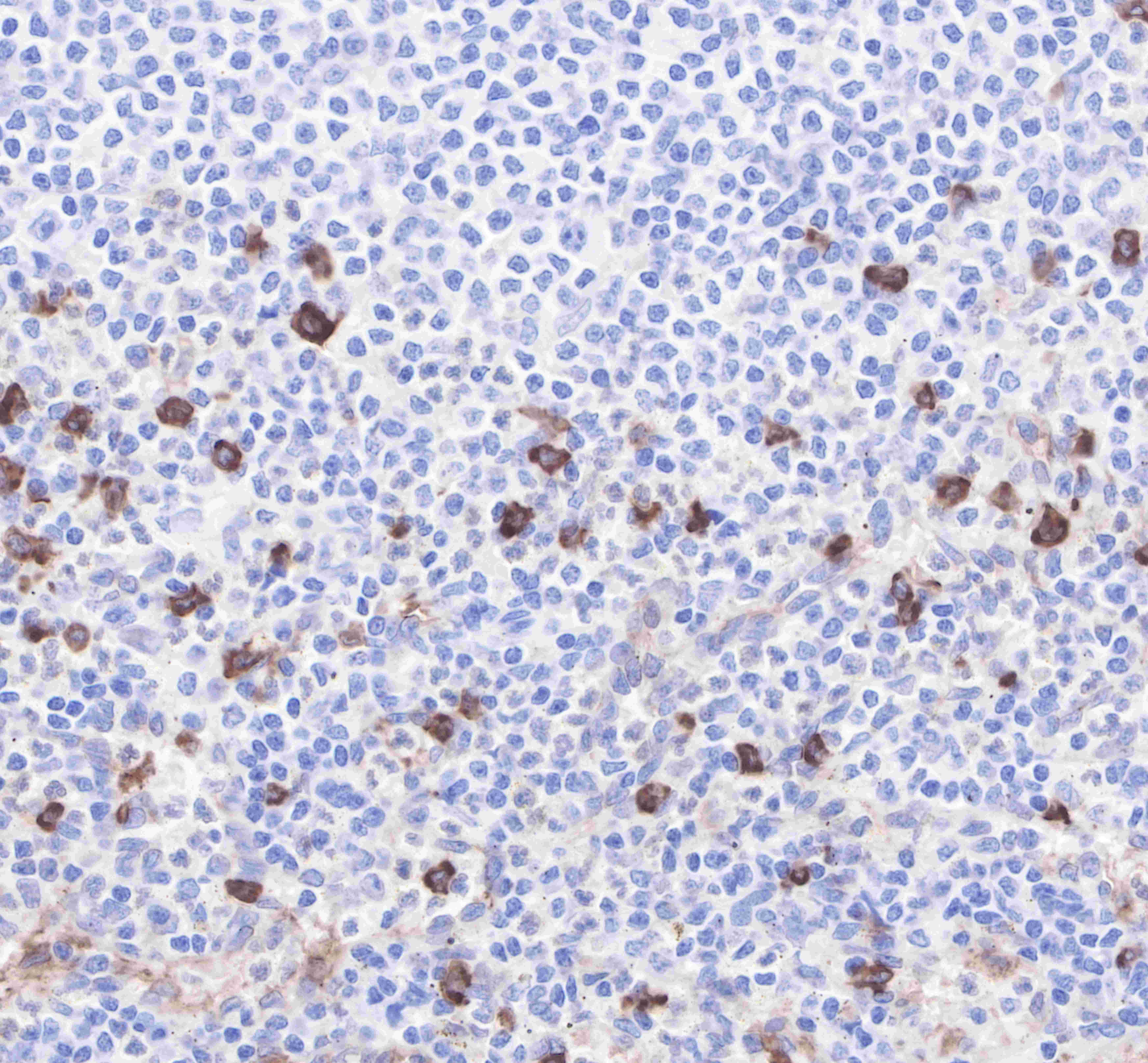
IHC shows positive staining in paraffin-embedded human tonsil. Anti-CD14 antibody was used at 1/2000 dilution, followed by a Goat Anti-Rabbit IgG H&L (HRP) ready to use.
Counterstained with hematoxylin.
Heat mediated antigen retrieval with Tris/EDTA buffer pH9.0 was performed before commencing with IHC staining protocol.
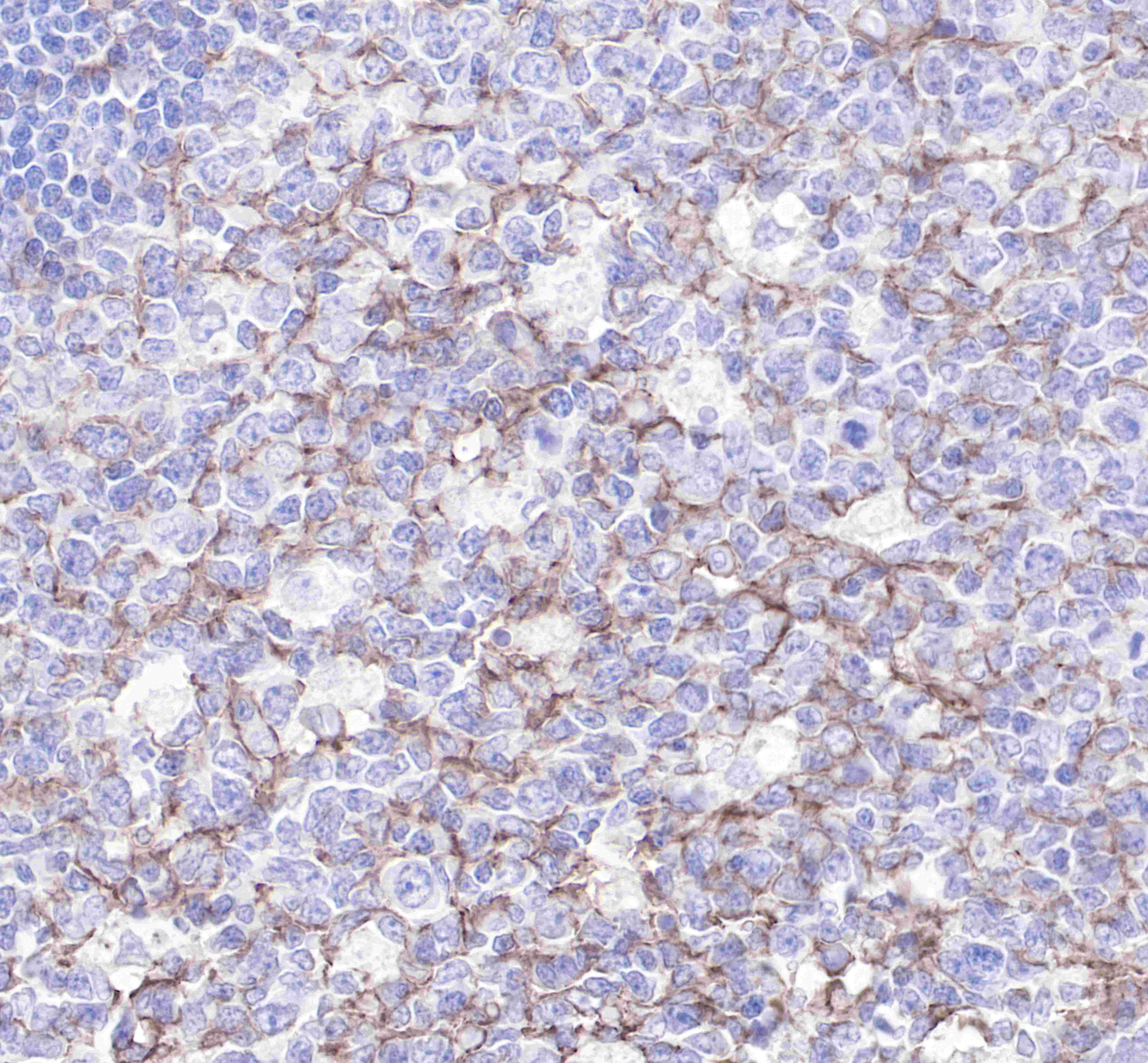
HC shows positive staining in paraffin-embedded human spleen. Anti-CD14 antibody was used at 1/2000 dilution, followed by a Goat Anti-Rabbit IgG H&L (HRP) ready to use.
Counterstained with hematoxylin.
Heat mediated antigen retrieval with Tris/EDTA buffer pH9.0 was performed before commencing with IHC staining protocol.
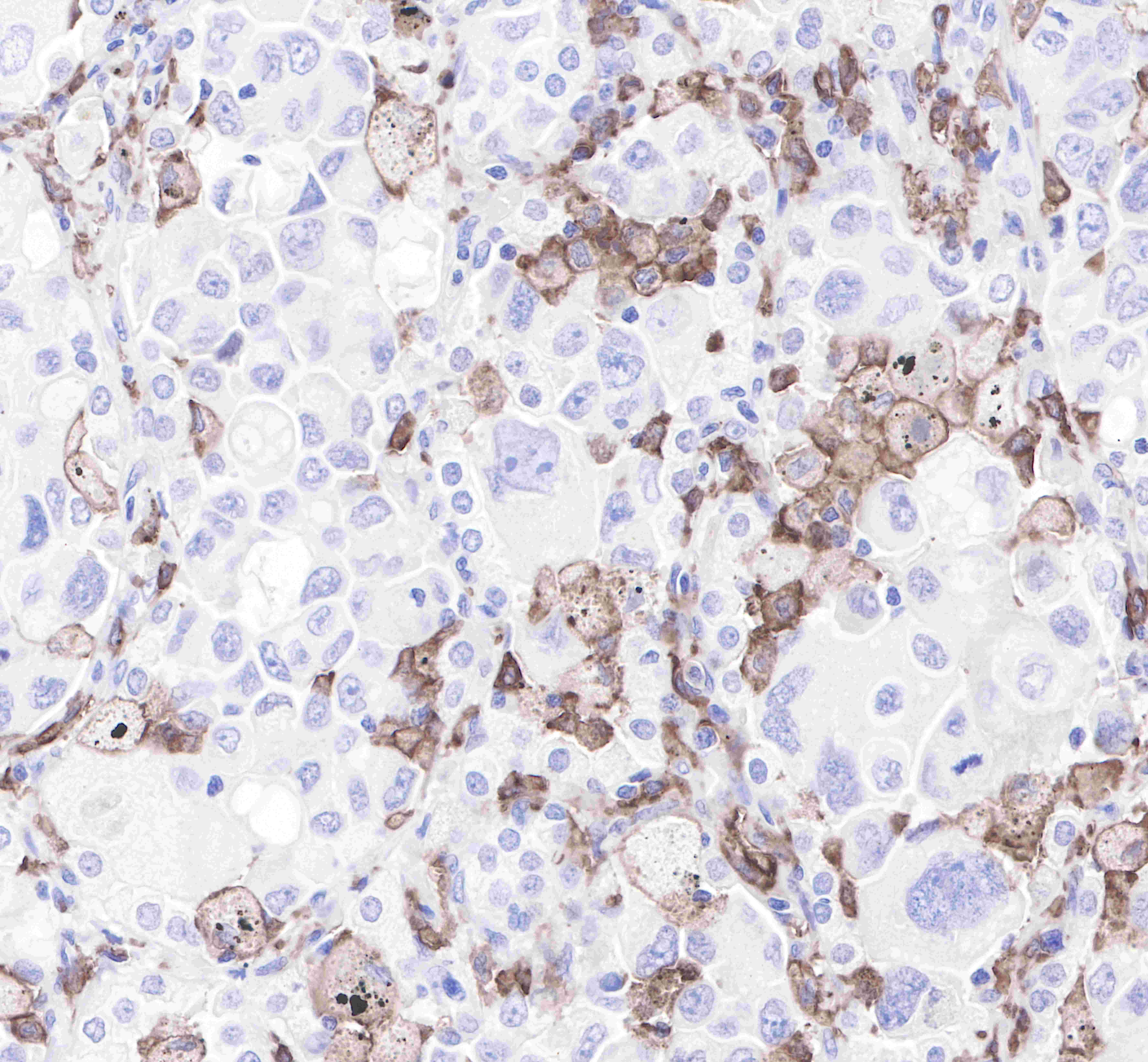
IHC shows positive staining in paraffin-embedded human stomach. Anti-CD14 antibody was used at 1/2000 dilution, followed by a Goat Anti-Rabbit IgG H&L (HRP) ready to use.
Counterstained with hematoxylin.
Heat mediated antigen retrieval with Tris/EDTA buffer pH9.0 was performed before commencing with IHC staining protocol.
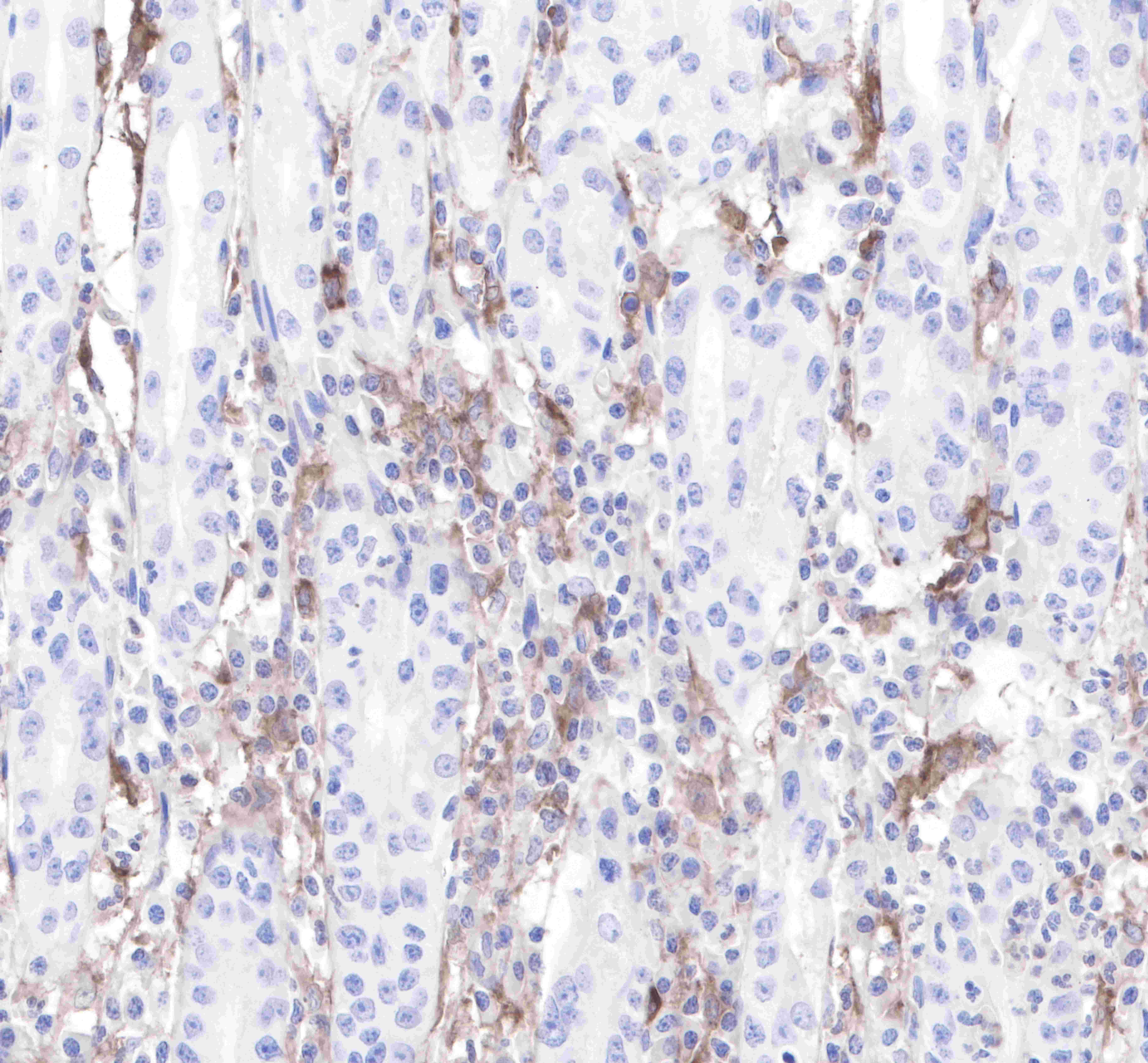
IHC shows positive staining in paraffin-embedded human ovarian cancer. Anti-CD14 antibody was used at 1/2000 dilution, followed by a Goat Anti-Rabbit IgG H&L (HRP) ready to use.
Counterstained with hematoxylin.
Heat mediated antigen retrieval with Tris/EDTA buffer pH9.0 was performed before commencing with IHC staining protocol.
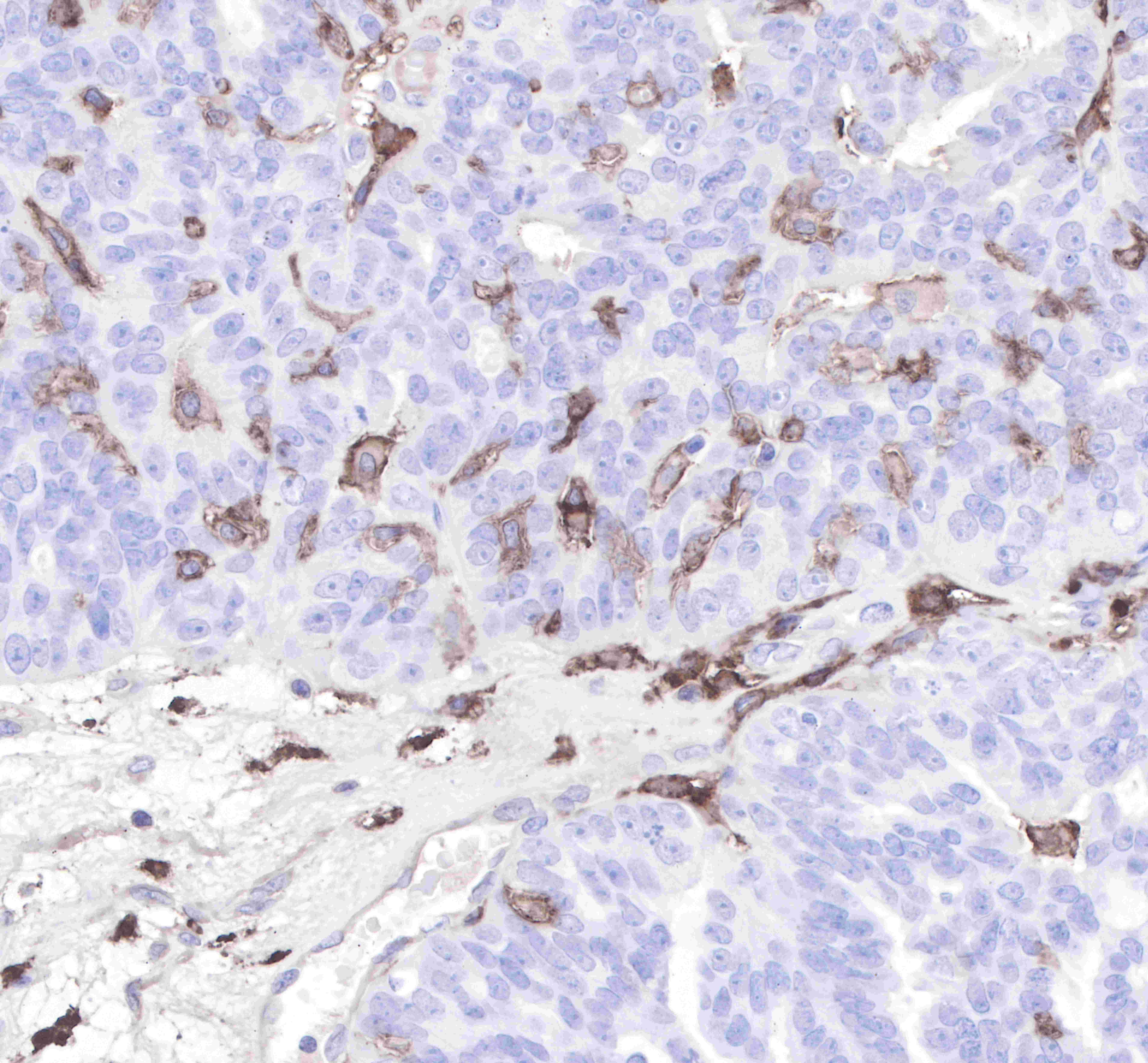
IHC shows positive staining in paraffin-embedded human lung adenocarcinoma.
Anti-CD14 antibody was used at 1/2000 dilution, followed by a Goat Anti-Rabbit IgG H&L (HRP) ready to use.
Counterstained with hematoxylin.
Heat mediated antigen retrieval with Tris/EDTA buffer pH9.0 was performed before commencing with IHC staining protocol.
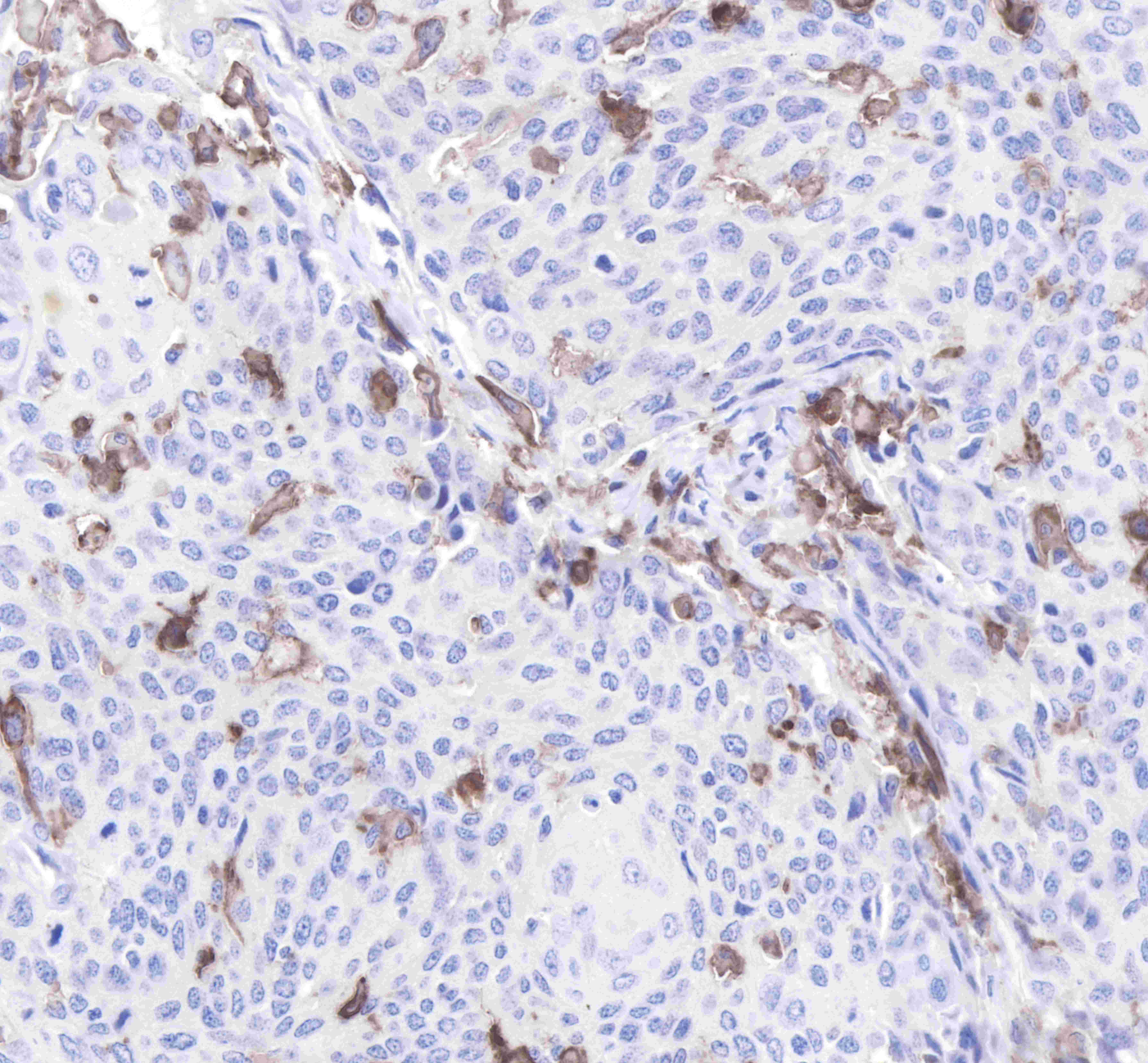
IHC shows positive staining in paraffin-embedded human cervix cancer.
Anti-CD14 antibody was used at 1/2000 dilution, followed by a Goat Anti-Rabbit IgG H&L (HRP) ready to use.
Counterstained with hematoxylin.
Heat mediated antigen retrieval with Tris/EDTA buffer pH9.0 was performed before commencing with IHC staining protocol.

ICC shows positive staining in THP-1 cells. Anti-CD14 antibody was used at 1/500 dilution (Green) and incubated overnight at 4°C. Goat polyclonal Antibody to Rabbit IgG - H&L (Alexa Fluor® 488) was used as secondary antibody at 1/1000 dilution. The cells were fixed with 100% ice-cold methanol and permeabilized with 0.1% PBS-Triton X-100. Nuclei were counterstained with DAPI (Blue).Counterstain with tubulin (red).
 您现在的位置: