
PBS, 40% Glycerol, 0.05%BSA, 0.03% Proclin 300
12 months from date of receipt / reconstitution, -20 °C as supplied
| 应用 | 稀释度 |
|---|---|
| ICC | 1:500 |
| WB | 1:5000 |
| ICFCM | 1:500 |
| IHC-P | 1:500 |
| IP | 1:50 |
Enhancer of zeste homolog 2 (EZH2) is a histone-lysine N-methyltransferase enzyme (EC 2.1.1.43) encoded by EZH2 gene, that participates in histone methylation and, ultimately, transcriptional repression. EZH2 catalyzes the addition of methyl groups to histone H3 at lysine 27, by using the cofactor S-adenosyl-L-methionine. Methylation activity of EZH2 facilitates heterochromatin formation thereby silences gene function. Remodeling of chromosomal heterochromatin by EZH2 is also required during cell mitosis.

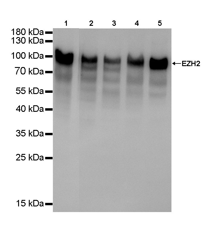
WB result of EZH2 Rabbit mAb
Primary antibody: EZH2 Rabbit mAb at 1/5000 dilution
Lane 1: Jurkat whole cell lysate 20 µg
Lane 2: T47D whole cell lysate 20 µg
Lane 3: MCF7 whole cell lysate 20 µg
Lane 4: Hela whole cell lysate 20 µg
Lane 5: neuro-2a whole cell lysate 20 µg
Secondary antibody: Goat Anti-Rabbit IgG, (H+L), HRP conjugated at 1/10000 dilution
Predicted MW: 100 kDa
Observed MW: 100 kDa
Exposure time: Lane 1: 15s
Lane 2~Lane 5: 20s
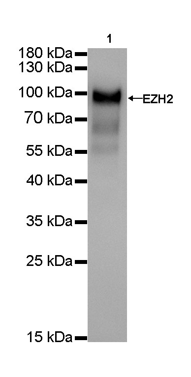
WB result of EZH2 Rabbit mAb
Primary antibody: EZH2 Rabbit mAb at 1/5000 dilution
Lane 1: NIH/3T3 whole cell lysate 20 µg
Secondary antibody: Goat Anti-Rabbit IgG, (H+L), HRP conjugated at 1/10000 dilution
Predicted MW: 100 kDa
Observed MW: 100 kDa
Exposure time: 20s
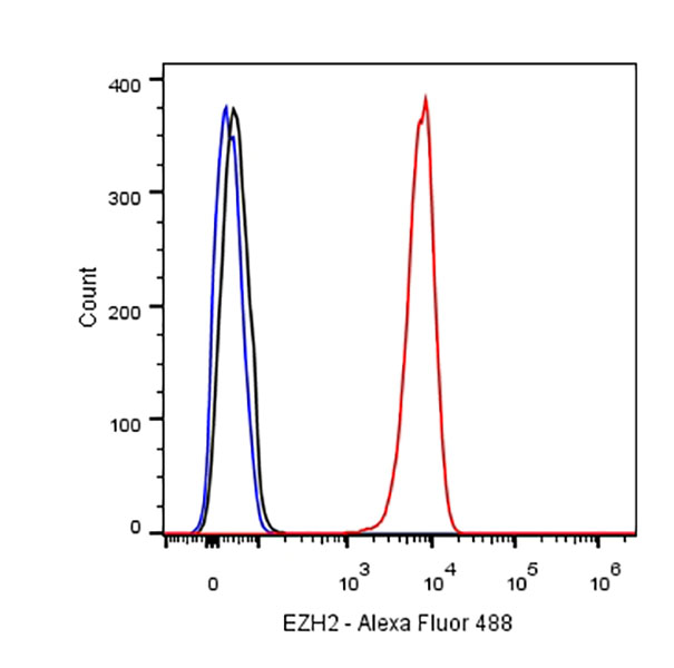
Flow cytometric analysis of Jurkat cells labelling EZH2 antibody at 1/500 (0.1ug) dilution/ (red) compared with a Rabbit monoclonal IgG (Black) isotype control and an unlabelled control (cells without incubation with primary antibody and secondary antibody) (Blue). Goat Anti-Rabbit IgG Alexa Fluor® 488 was used as the secondary antibody.
EZH2 Rabbit mAb at 1/50 dilution (1 µg) immunoprecipitating EZH2 in 0.4 mg Jurkat whole cell lysate.
Western blot was performed on the immunoprecipitate using EZH2 Rabbit mAb at 1/1000 dilution.
Secondary antibody (HRP) for IP was used at 1/400 dilution.
Lane 1: Jurkat whole cell lysate 10 µg (Input)
Lane 2: EZH2 Rabbit mAb IP in Jurkat whole cell lysate
Lane 3: Rabbit monoclonal IgG IP in Jurkat whole cell lysate
Predicted MW: 100 kDa
Observed MW: 100 kDa
Exposure time: 15 s
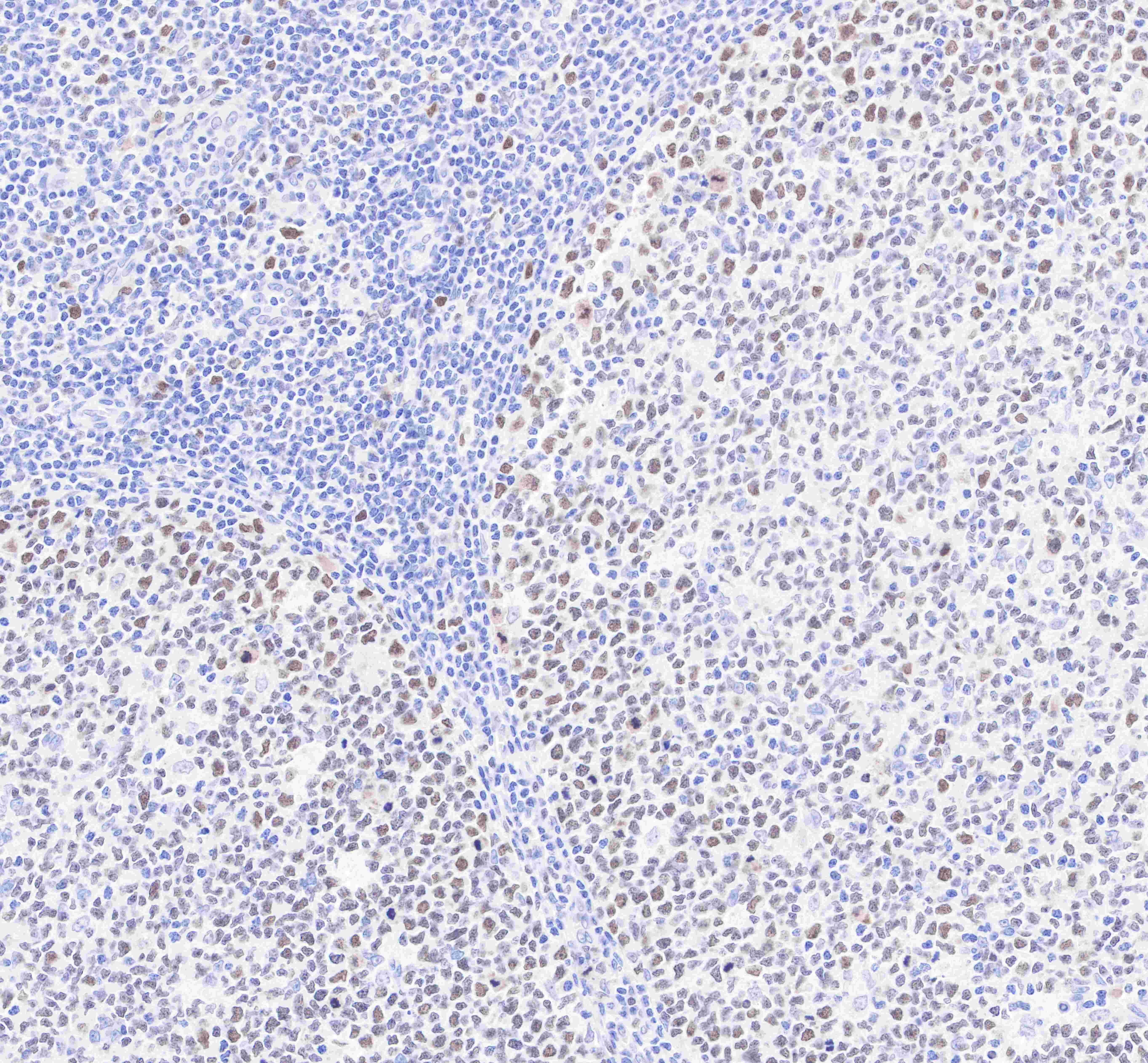
IHC shows positive staining in paraffin-embedded human tonsil.
Anti-EZH2 antibody was used at 1/500 dilution, followed by a Goat Anti-Rabbit IgG H&L (HRP) ready to use. Counterstained with hematoxylin.
Heat mediated antigen retrieval with Tris/EDTA buffer pH9.0 was performed before commencing with IHC staining protocol.
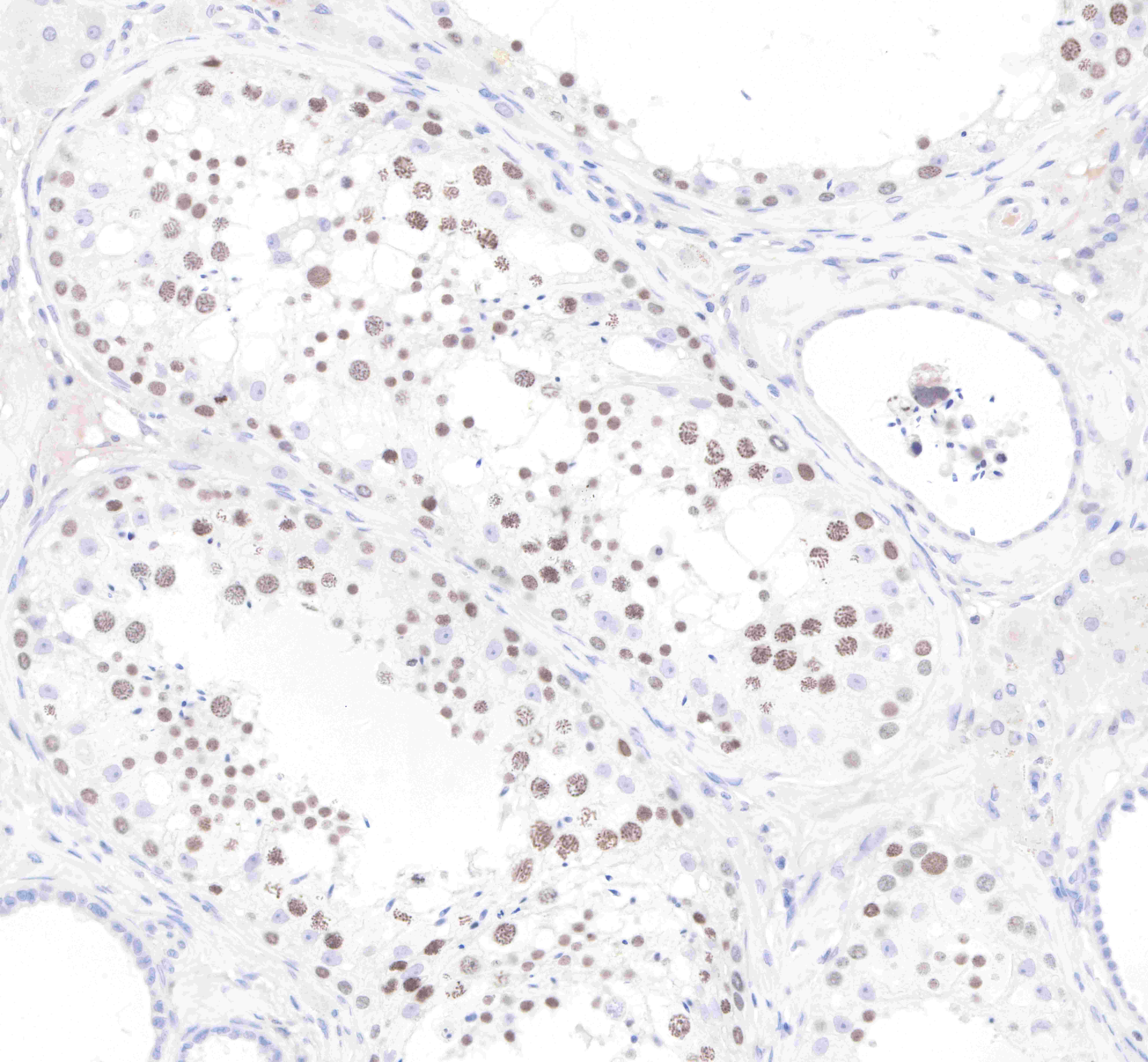
IHC shows positive staining in paraffin-embedded human testis.
Anti-EZH2 antibody was used at 1/500 dilution, followed by a Goat Anti-Rabbit IgG H&L (HRP) ready to use. Counterstained with hematoxylin.
Heat mediated antigen retrieval with Tris/EDTA buffer pH9.0 was performed before commencing with IHC staining protocol.
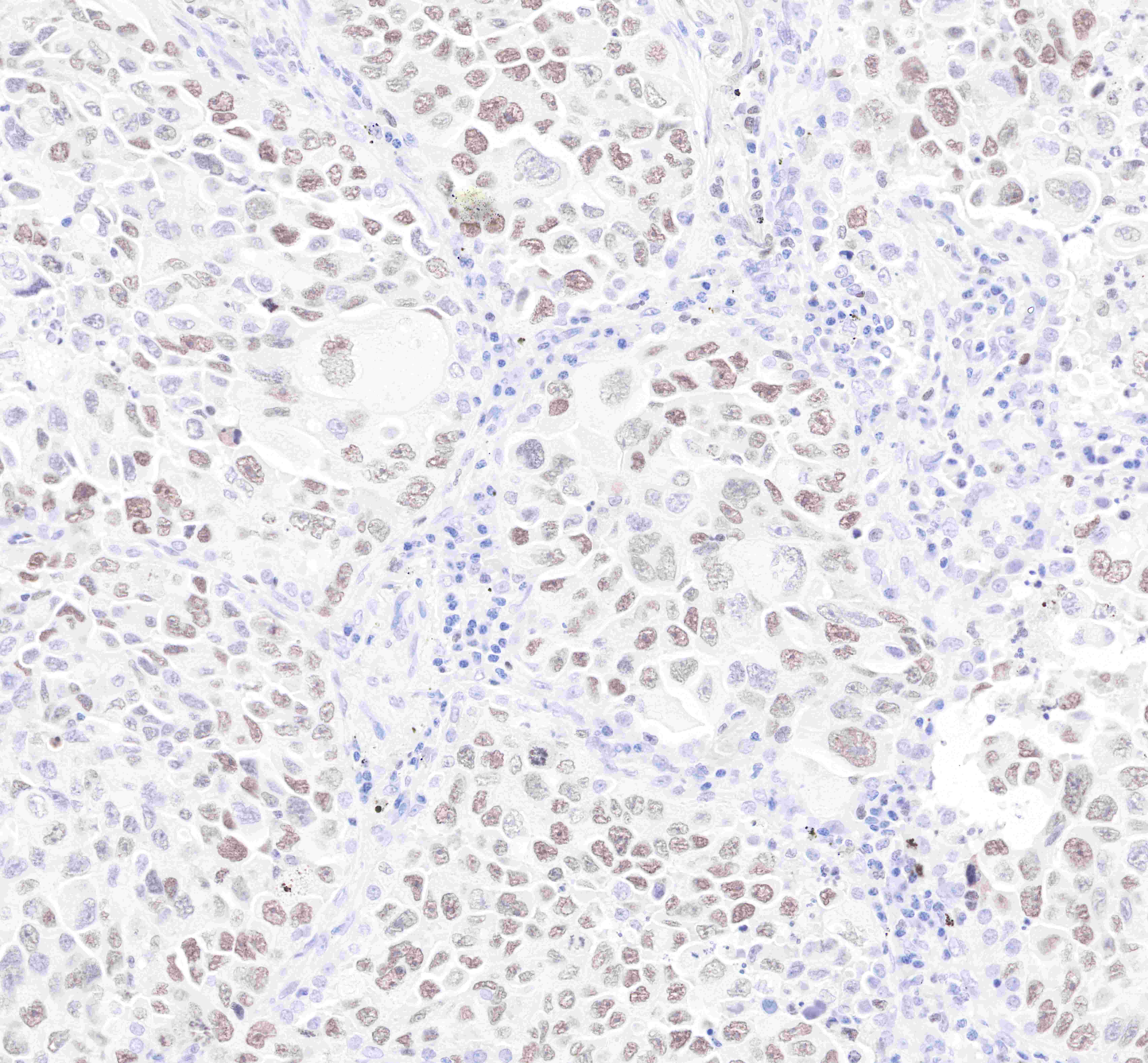
IHC shows positive staining in paraffin-embedded human lung adenocarcinoma.
Anti-EZH2 antibody was used at 1/500 dilution, followed by a Goat Anti-Rabbit IgG H&L (HRP) ready to use. Counterstained with hematoxylin.
Heat mediated antigen retrieval with Tris/EDTA buffer pH9.0 was performed before commencing with IHC staining protocol.
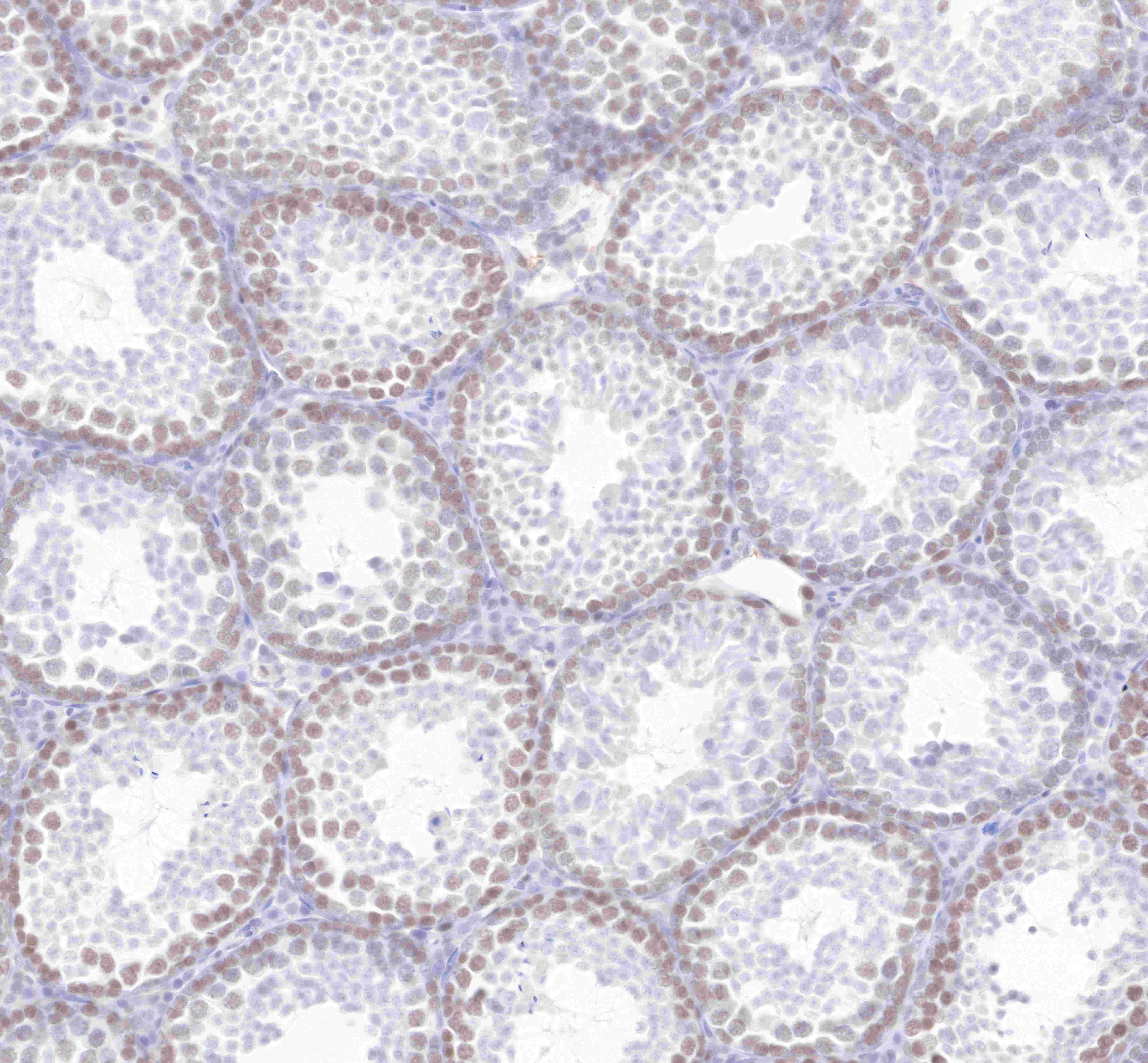
IHC shows positive staining in paraffin-embedded mouse testis.
Anti-EZH2 antibody was used at 1/500 dilution, followed by a Goat Anti-Rabbit IgG H&L (HRP) ready to use. Counterstained with hematoxylin.
Heat mediated antigen retrieval with Tris/EDTA buffer pH9.0 was performed before commencing with IHC staining protocol.

ICC shows positive staining in Jurkat cells. Anti-EZH2 antibody was used at 1/500 dilution (Green) and incubated overnight at 4°C. Goat polyclonal Antibody to Rabbit IgG - H&L (Alexa Fluor® 488) was used as secondary antibody at 1/1000 dilution. The cells were fixed with 4% PFA and permeabilized with 0.1% PBS-Triton X-100. Nuclei were counterstained with DAPI (Blue).Counterstain with tubulin (Red).
 您现在的位置: