
PBS, 40% Glycerol, 0.05%BSA, 0.03% Proclin 300
12 months from date of receipt / reconstitution, -20 °C as supplied
| 应用 | 稀释度 |
|---|---|
| IHC-P | 1:1000-1:4000 |
| ICFCM | 1:500 |
| WB | 1:1000 |
| ICC | 1:1000 |
Keratin 5, also known as KRT5, K5, or CK5, is a protein that is encoded in humans by the KRT5 gene. It dimerizes with keratin 14 and forms the intermediate filaments (IF) that make up the cytoskeleton of basal epithelial cells. Keratin 5 serves as a biomarker for several different types of cancer, including breast and lung cancers. It is often tested in conjunction with keratin 6, using CK5/6 antibodies, which target both keratin forms.

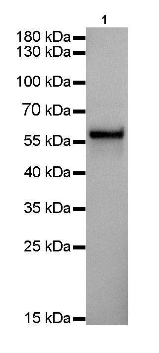
WB result of Keratin5 Rabbit mAb
Primary antibody: Keratin5 Rabbit mAb at 1/1000 dilution
Lane 1: A431 whole cell lysate 20 µg
Secondary antibody: Goat Anti-Rabbit IgG, (H+L), HRP conjugated at 1/10000 dilution
Predicted MW: 62 kDa
Observed MW: 62 kDa
Exposure time: 0.2s
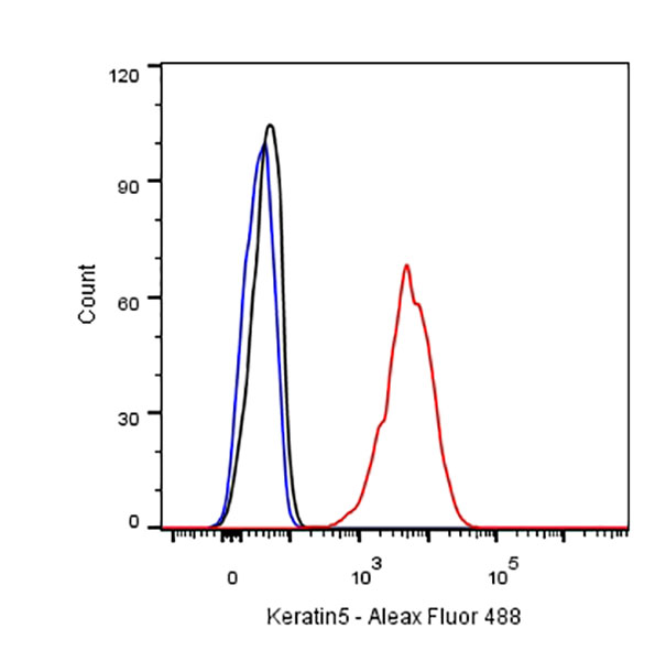
Flow cytometric analysis of A431 cells labelling Keratin5 antibody at 1/500 (0.1ug) dilution/ (red) compared with a Rabbit monoclonal IgG (Black) isotype control and an unlabelled control (cells without incubation with primary antibody and secondary antibody) (Blue). Goat Anti-Rabbit IgG Alexa Fluor® 488 was used as the secondary antibody.
IHC shows positive staining in paraffin-embedded human tonsil.
Anti-Keratin 5 antibody was used at 1/4000 dilution, followed by a Goat Anti-Rabbit IgG H&L (HRP) ready to use.
Counterstained with hematoxylin.
Heat mediated antigen retrieval with Tris/EDTA buffer pH9.0 was performed before commencing with IHC staining protocol.
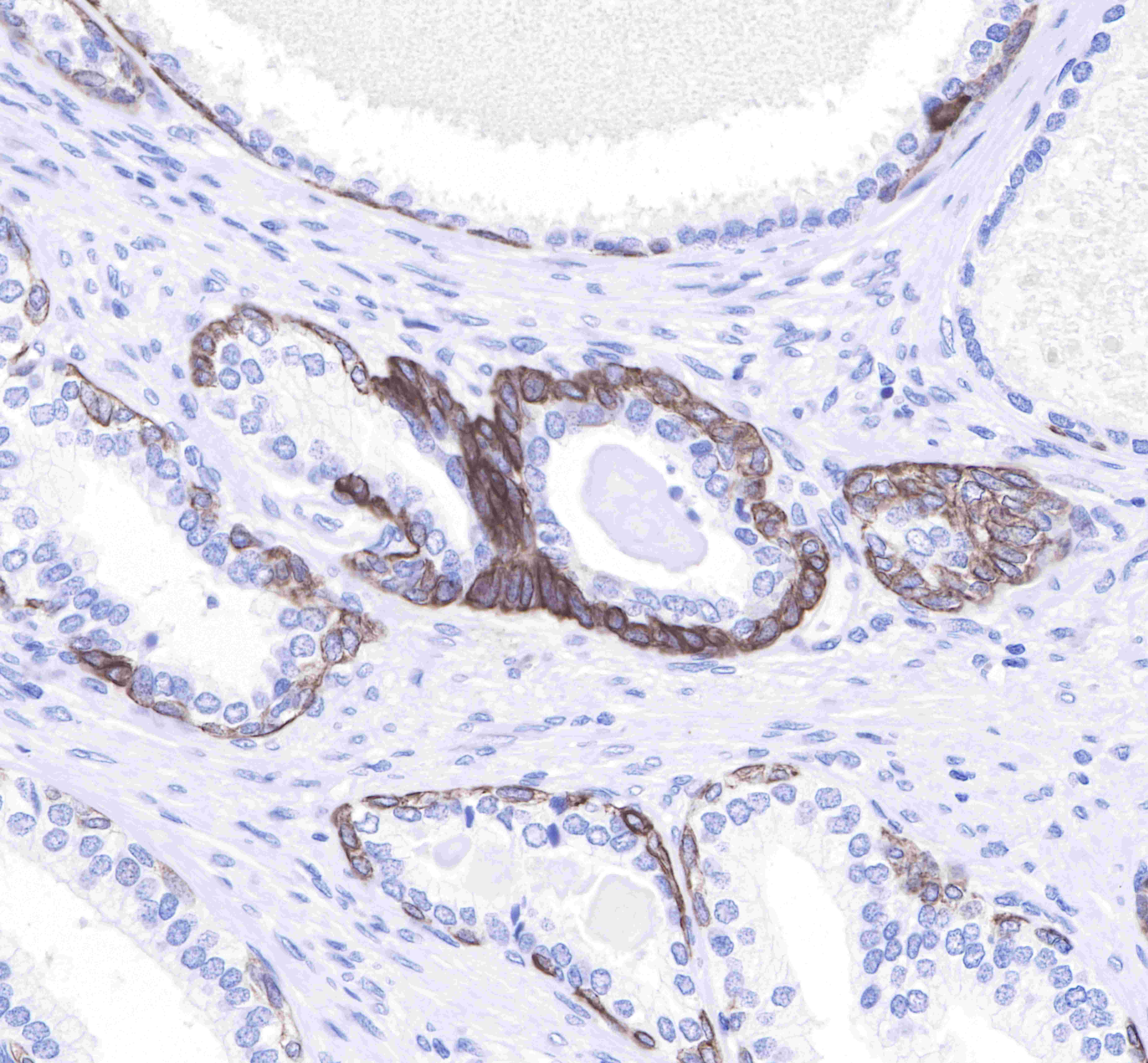
IHC shows positive staining in paraffin-embedded human prostate.
Anti-Keratin 5 antibody was used at 1/4000 dilution, followed by a Goat Anti-Rabbit IgG H&L (HRP) ready to use.
Counterstained with hematoxylin.
Heat mediated antigen retrieval with Tris/EDTA buffer pH9.0 was performed before commencing with IHC staining protocol.
Negative control: IHC shows negative staining in paraffin-embedded human colon.
Anti-Keratin 5 antibody was used at 1/4000 dilution, followed by a Goat Anti-Rabbit IgG H&L (HRP) ready to use.
Counterstained with hematoxylin.
Heat mediated antigen retrieval with Tris/EDTA buffer pH9.0 was performed before commencing with IHC staining protocol.
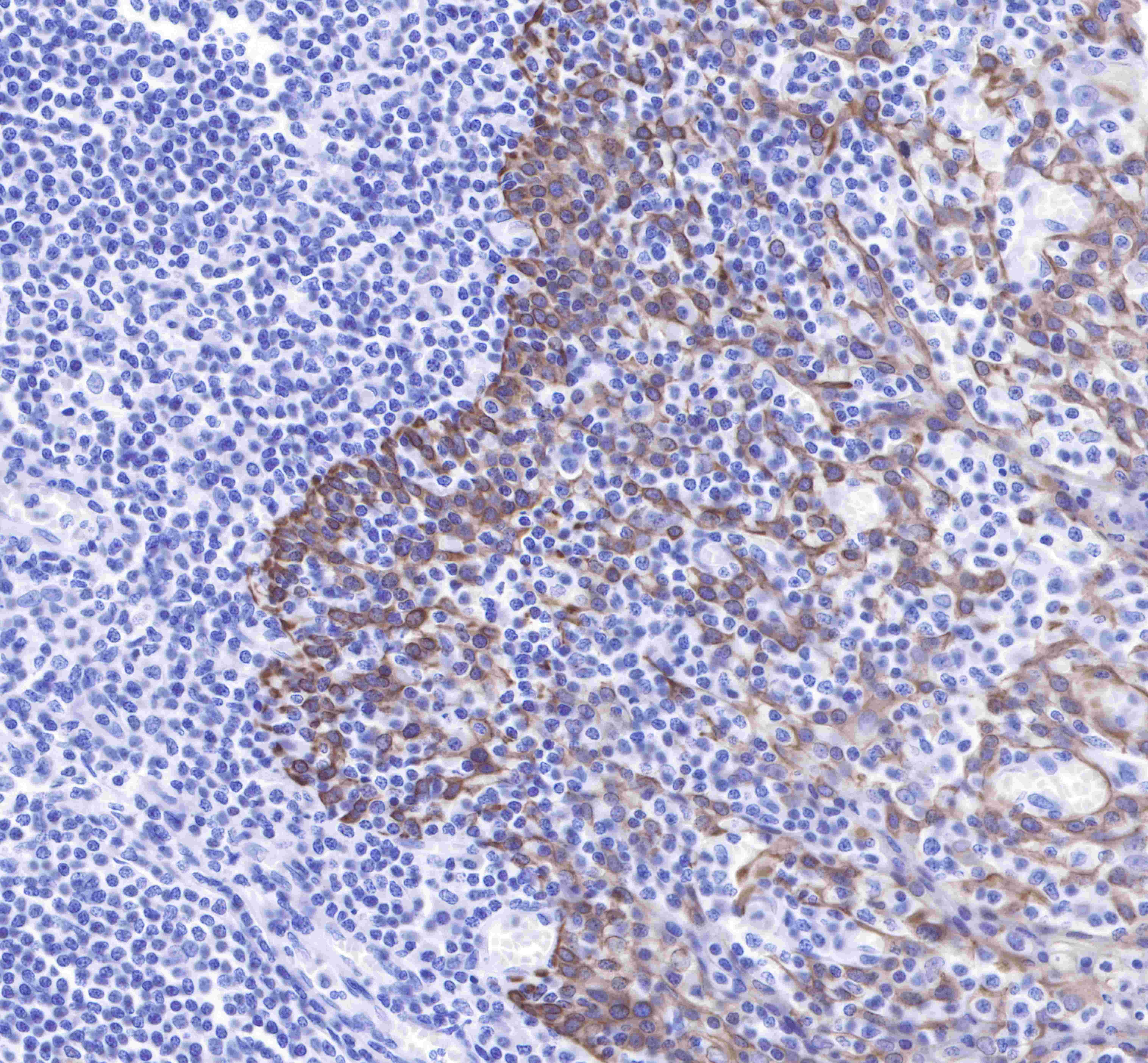
IHC shows positive staining in paraffin-embedded human lung squamous cell cancer.
Anti-Keratin 5 antibody was used at 1/4000 dilution, followed by a Goat Anti-Rabbit IgG H&L (HRP) ready to use.
Counterstained with hematoxylin.
Heat mediated antigen retrieval with Tris/EDTA buffer pH9.0 was performed before commencing with IHC staining protocol.
Negative control: IHC shows negative staining in paraffin-embedded human lung adenocarcinoma.
Anti-Keratin 5 antibody was used at 1/4000 dilution, followed by a Goat Anti-Rabbit IgG H&L (HRP) ready to use.
Counterstained with hematoxylin.
Heat mediated antigen retrieval with Tris/EDTA buffer pH9.0 was performed before commencing with IHC staining protocol.
ICC shows cytoplasm staining in A431 cells.
Anti-Keratin antibody was used at 1/1000 dilution and incubated overnight at 4°C.
Goat polyclonal Antibody to Rabbit IgG - H&L (Alexa Fluor® 488) was used as secondary antibody at 1/1000 dilution.
The cells were fixed with 100% Methonal and permeabilized with 0.1% PBS-Triton X-100.
Nuclei were counterstained with DAPI.
 您现在的位置: