

| 应用 | 稀释度 |
|---|---|
| IHC-P | 1:500 |
| ICC | 1:100 |
| WB | 1:1000 |
| IP | 1:50 |
T-cell surface glycoprotein CD3 gamma chain is a protein that in humans is encoded by the CD3G gene. T cell antigen receptor (TCR) is associated on the T cell surface with a complex of protein called CD3. CD3G (gamma chain) is one of the four peptides (gamma, delta, epsilon and zeta) that form CD3. Defects in CD3G are associated with T cell immunodeficiency.
WB result of CD3G Rabbit mAb
Primary antibody: CD3G Rabbit mAb at 1/1000 dilution
Lane 1: Jurkat whole cell lysate 20 µg
Secondary antibody: Goat Anti-Rabbit IgG, (H+L), HRP conjugated at 1/10000 dilution
Predicted MW: 20 kDa
Observed MW: 20 kDa
Exposure time: 10s

CD3G Rabbit mAb at 1/50 dilution (1 µg) immunoprecipitating CD3G in 0.4 mg Jurkat whole cell lysate.
Western blot was performed on the immunoprecipitate using CD3G Rabbit mAb at 1/1000 dilution.
Secondary antibody (HRP) for IP was used at 1/400 dilution.
Lane 1: Jurkat whole cell lysate 30 µg (Input)
Lane 2: CD3G Rabbit mAb IP in Jurkat whole cell lysate
Lane 3: Rabbit monoclonal IgG IP in Jurkat whole cell lysate
Predicted MW: 20 kDa
Observed MW: 20 kDa
Exposure time: 60 s
IHC shows positive staining in paraffin-embedded human tonsil.
Anti-CD3G antibody was used at 1/500 dilution, followed by a Goat Anti-Rabbit IgG H&L (HRP) ready to use.
Counterstained with hematoxylin.
Heat mediated antigen retrieval with Tris/EDTA buffer pH9.0 was performed before commencing with IHC staining protocol.
IHC shows positive staining in paraffin-embedded human spleen.
Anti-CD3G antibody was used at 1/500 dilution, followed by a Goat Anti-Rabbit IgG H&L (HRP) ready to use.
Counterstained with hematoxylin.
Heat mediated antigen retrieval with Tris/EDTA buffer pH9.0 was performed before commencing with IHC staining protocol.
IHC shows positive staining in paraffin-embedded human colon.
Anti-CD3G antibody was used at 1/500 dilution, followed by a Goat Anti-Rabbit IgG H&L (HRP) ready to use.
Counterstained with hematoxylin.
Heat mediated antigen retrieval with Tris/EDTA buffer pH9.0 was performed before commencing with IHC staining protocol.
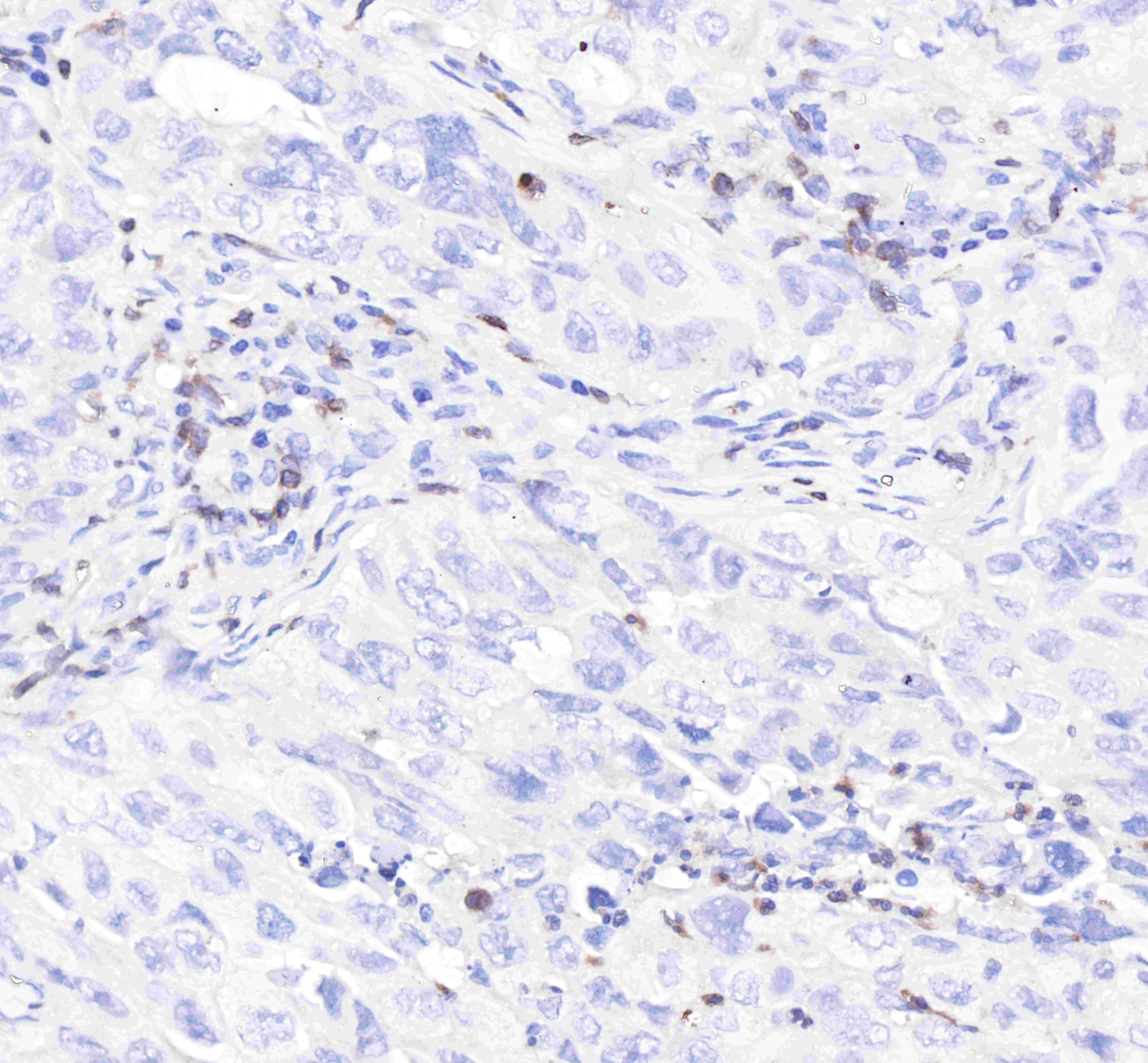
IHC shows positive staining in paraffin-embedded human lung cancer.
Anti-CD3G antibody was used at 1/500 dilution, followed by a Goat Anti-Rabbit IgG H&L (HRP) ready to use.
Counterstained with hematoxylin.
Heat mediated antigen retrieval with Tris/EDTA buffer pH9.0 was performed before commencing with IHC staining protocol.
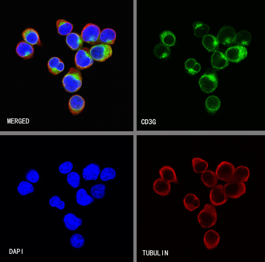
ICC shows positive staining in Jurkat cells. Anti-CD3G antibody was used at 1/100 dilution (Green) and incubated overnight at 4°C. Goat polyclonal Antibody to Rabbit IgG - H&L (Alexa Fluor® 488) was used as secondary antibody at 1/1000 dilution. The cells were fixed with 4% PFA and permeabilized with 0.1% PBS-Triton X-100. Nuclei were counterstained with DAPI (Blue). Counterstain with tubulin (Red).
 您现在的位置: