

| 应用 | 稀释度 |
|---|---|
| IP | 1:25 |
| IHC-P | 1:500-2000 |
| WB | 1:1000 |
| ICC | 1:500 |
| ICFCM | 1:5000 |
Aurora kinase B is a protein that functions in the attachment of the mitotic spindle to the centromere. The expression and activity of Aurora B are regulated according to the cell cycle. Expression of Aurora B reaches a maximum at the G2-M transition, whereas Aurora B protein is most active during mitosis. The Aurora kinases associate with microtubules during chromosome movement and segregation. Aurora kinase B localizes to microtubules near kinetochores, specifically to the specialized microtubules called K-fibers, and Aurora kinase A localizes to centrosomes. In cancerous cells, over-expression of these enzymes causes unequal distribution of genetic information, creating aneuploid cells, a hallmark of cancer.
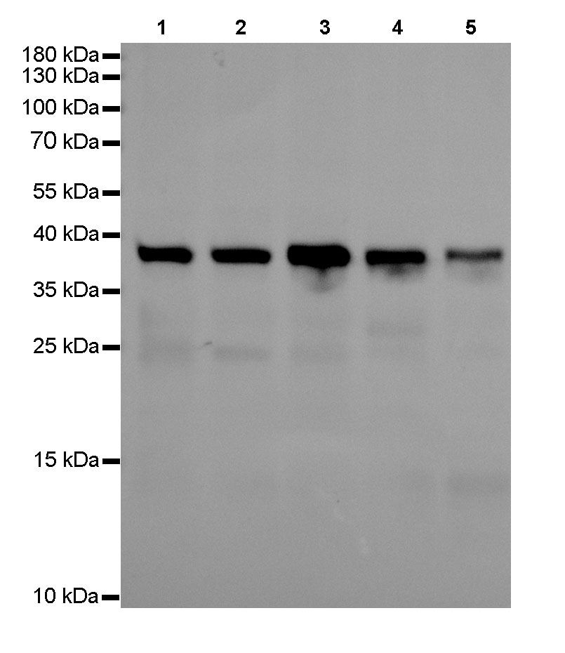
WB result of Aurora-B Rabbit mAb
Primary antibody : Aurora-B Rabbit mAb at 1/1000 dilution
Lane 1: Hela whole cell lysate 20 µg
Lane 2: HepG2 whole cell lysate 20 µg
Lane 3: Raji whole cell lysate 20 µg
Lane 4: T47D whole cell lysate 20 µg
Lane 5: A549 whole cell lysate 20 µg
Secondary antibody: Goat Anti-Rabbit IgG, (H+L), HRP conjugated at 1/10000 dilution
Predicted MW: 41 kDa
Observed MW: 39 kDa
Exposure time: 16 s
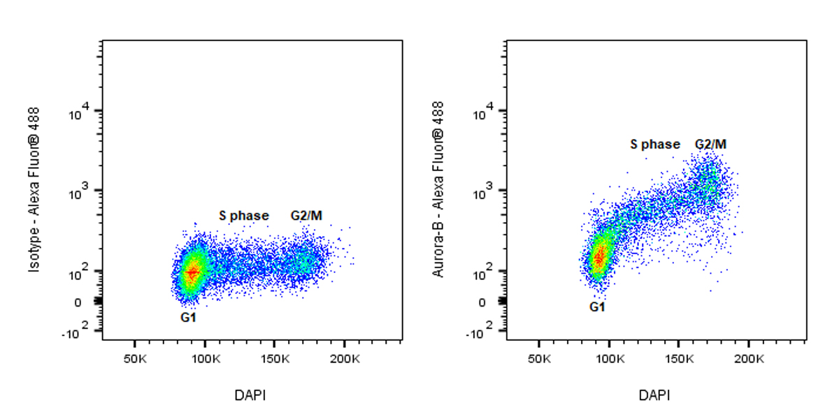
Flow cytometric analysis of 4% PFA fixed 90% methanol permeabilized HeLa (Human cervix adenocarcinoma epithelial cell) labelling Aurora-B antibody at 1/5000 dilution (0.01 μg)/ (Right) compared with a Rabbit monoclonal IgG / (Left) isotype control. Goat Anti - Rabbit IgG Alexa Fluor® 488 was used as the secondary antibody. Cells were co-stained with DAPI to differentiate cell cycle phase.
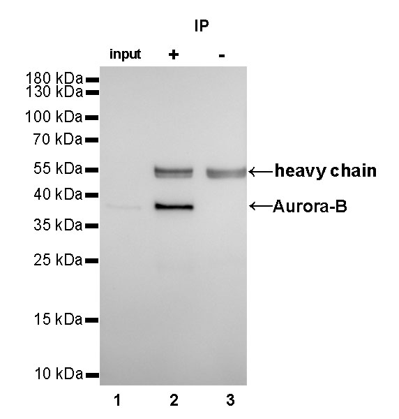
Aurora-B Rabbit mAb at 1/25 dilution (2µg) immunoprecipitating Aurora-B in 0.4mg HeLa whole cell lysate.
Western blot was performed on the immunoprecipitate using Aurora-B Rabbit mAb at 1/1000 dilution.
Secondary antibody (HRP) for IP was used at 1/400 dilution.
Lane 1 : HeLa whole cell lysate 10µg (input)
Lane 2 (+): Aurora-B Rabbit mAb IP in HeLa whole cell lysate
Lane 3 (-): Rabbit monoclonal IgG IP in HeLa whole cell lysate
Predicted MW: 41 kDa
Observed MW: 39 kDa
Exposure time: 10s
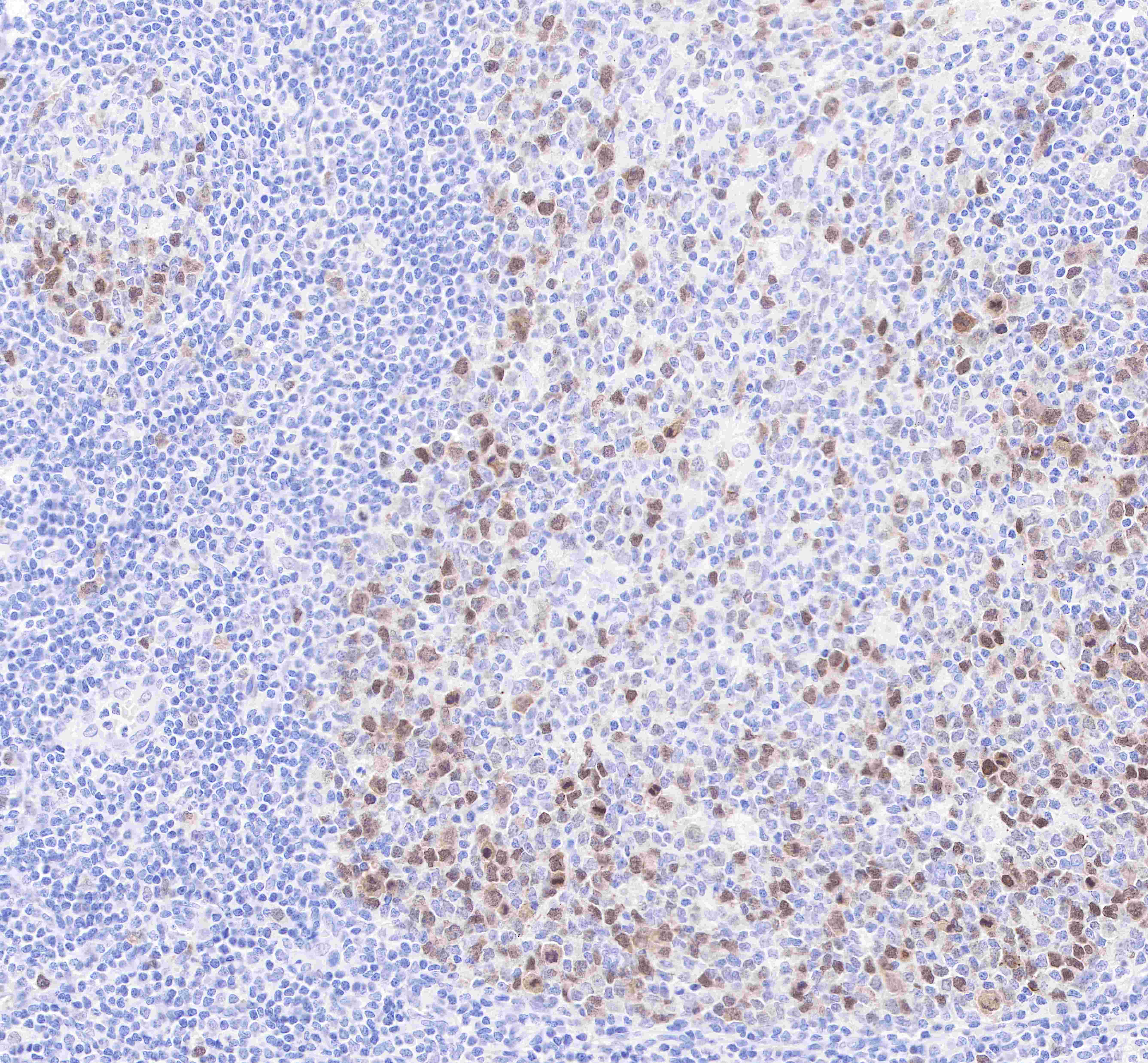
IHC shows positive staining in paraffin-embedded human tonsil.
Anti-Aurora B antibody was used at 1/2000 dilution, followed by a Goat Anti-Rabbit IgG H&L (HRP) ready to use.
Counterstained with hematoxylin.
Heat mediated antigen retrieval with Tris/EDTA buffer pH9.0 was performed before commencing with IHC staining protocol.

IHC shows positive staining in paraffin-embedded human cervix cancer.
Anti-Aurora B antibody was used at 1/500 dilution, followed by a Goat Anti-Rabbit IgG H&L (HRP) ready to use.
Counterstained with hematoxylin.
Heat mediated antigen retrieval with Tris/EDTA buffer pH9.0 was performed before commencing with IHC staining protocol.
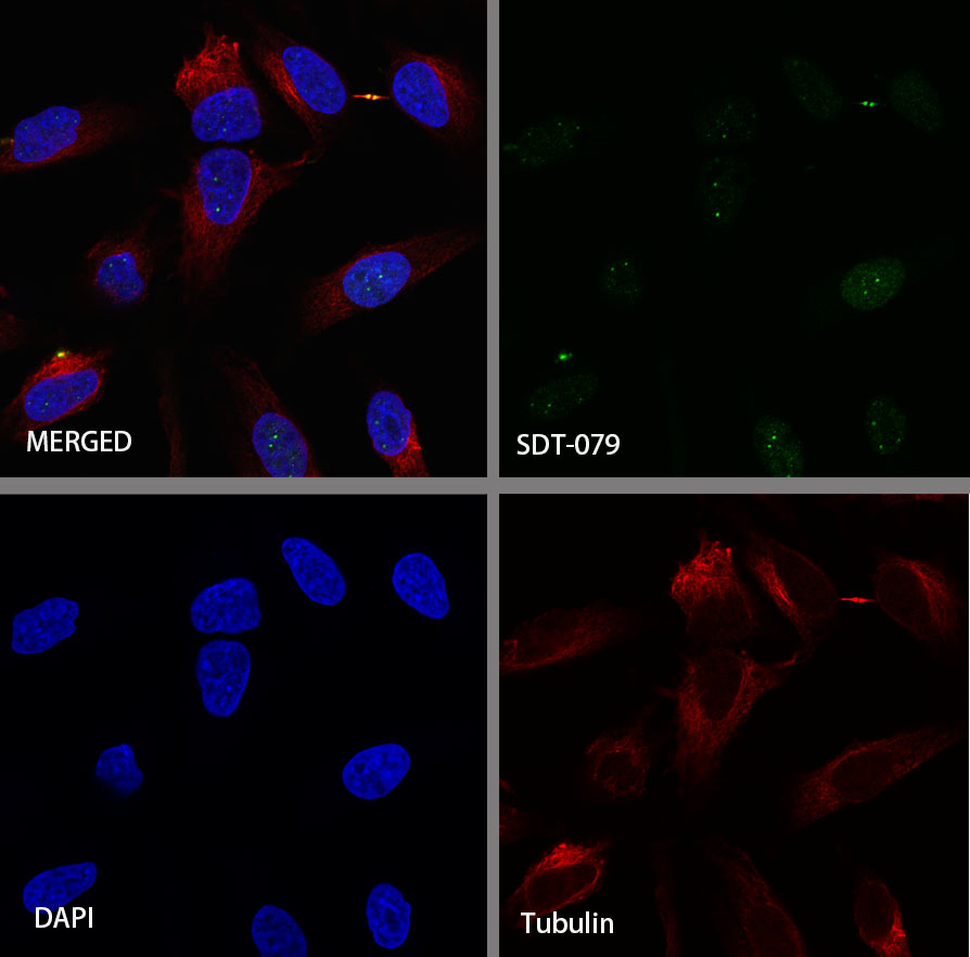
ICC shows positive staining in HeLa cells. Anti-Aurora-B antibody was used at 1/500 dilution (Green) and incubated overnight at 4°C. Goat polyclonal Antibody to Rabbit IgG - H&L (Alexa Fluor® 488) was used as secondary antibody at 1/1000 dilution. The cells were fixed with 4%PFA and permeabilized with 0.1% PBS-Triton X-100. Nuclei were counterstained with DAPI (Blue).Counterstain with tubulin (red).
 您现在的位置: