
12 months from date of receipt / reconstitution, -20 °C as supplied
| 应用 | 稀释度 |
|---|---|
| WB | 1:1000 |
| IP | 1:50 |
| IHC-P | 1:250 |
| ICC | 1:500 |
c-IAP1, which stands for cellular Inhibitor of Apoptosis Protein 1, is a member of the Inhibitor of Apoptosis (IAP) family. These proteins are known for their anti-apoptotic properties and are involved in the regulation of cell survival and death pathways. c-IAP1, in particular, has been shown to inhibit apoptosis by directly binding to and inhibiting the activity of specific caspases, which are cysteine proteases that play a central role in the execution of apoptosis. Abnormal expression of IAP family members, including c-IAP1, has been associated with various cancers, suggesting their role in tumorigenesis and resistance to therapy. High expression levels of c-IAP1 in certain cancer cells have been linked to increased resistance to radiation and chemotherapy, making it a potential target for cancer therapy.
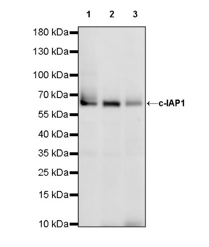
WB result of c-IAP1 Recombinant Rabbit mAb
Primary antibody: c-IAP1 Recombinant Rabbit mAb at 1/1000 dilution
Lane 1: HeLa whole cell lysate 20 µg
Lane 2: HepG2 whole cell lysate 20 µg
Lane 3: HT-29 whole cell lysate 20 µg
Secondary antibody: Goat Anti-rabbit IgG, (H+L), HRP conjugated at 1/10000 dilution
Predicted MW: 70 kDa
Observed MW: 65 kDa
This blot was developed with high sensitivity substrate
c-IAP1 Rabbit mAb at 1/50 dilution (1 µg) immunoprecipitating c-IAP1 in 0.4 mg HepG2 whole cell lysate.
Western blot was performed on the immunoprecipitate using c-IAP1 Rabbit mAb at 1/1000 dilution.
Secondary antibody (HRP) for IP was used at 1/1000 dilution.
Lane 1: HepG2 whole cell lysate 20 µg (Input)
Lane 2: c-IAP1 Rabbit mAb IP in HepG2 whole cell lysate
Lane 3: Rabbit monoclonal IgG IP in HepG2 whole cell lysate
Predicted MW: 70 kDa
Observed MW: 65 kDa
This blot was developed with high sensitivity substrate
IHC shows positive staining in paraffin-embedded human spleen. Anti- c-IAP1 antibody was used at 1/250 dilution, followed by a HRP Polymer for Mouse & Rabbit IgG (ready to use). Counterstained with hematoxylin. Heat mediated antigen retrieval with Tris/EDTA buffer pH9.0 was performed before commencing with IHC staining protocol.
IHC shows positive staining in paraffin-embedded human lung squamous cell carcinoma. Anti- c-IAP1 antibody was used at 1/250 dilution, followed by a HRP Polymer for Mouse & Rabbit IgG (ready to use). Counterstained with hematoxylin. Heat mediated antigen retrieval with Tris/EDTA buffer pH9.0 was performed before commencing with IHC staining protocol.
ICC shows positive staining in HepG2 cells. Anti-c-IAP1 antibody was used at 1/500 dilution (Green) and incubated overnight at 4°C. Goat polyclonal Antibody to Rabbit IgG - H&L (Alexa Fluor® 488) was used as secondary antibody at 1/1000 dilution. The cells were fixed with 100% ice-cold methanol and permeabilized with 0.1% PBS-Triton X-100. Nuclei were counterstained with DAPI (Blue). Counterstain with tubulin (Red).
 您现在的位置: