
PBS, 40% Glycerol, 0.05% BSA, 0.03% Proclin 300
12 months from date of receipt / reconstitution, -20 °C as supplied
| 应用 | 稀释度 |
|---|---|
| Dot Blot | 1:1000 |
| WB | 1:1000 |
| IHC-P | 1:200 |
| ICC | 1:500 |
| ChIP | 1:20~1:50 |
Acetylated histones are a key component of the epigenetic regulation of gene expression in eukaryotic cells. Histone acetylation involves the addition of an acetyl group to the lysine residues on the histone proteins that make up the nucleosome, which is the basic unit of chromatin. This process is generally associated with transcriptional activation, as it loosens the interaction between the DNA and the histones, allowing the transcription machinery to access the DNA and read the genetic information. Abnormalities in histone acetylation, such as global hypoacetylation or hyperacetylation, can lead to dysregulation of gene expression and have been implicated in various diseases, including cancer. Understanding the role of acetylated histones in gene regulation is crucial for developing therapeutic strategies for these diseases.
WB result of Histone H4 (acetyl K5 + K8 + K12 + K16) Recombinant Rabbit mAb
Primary antibody: Histone H4 (acetyl K5 + K8 + K12 + K16) Recombinant Rabbit mAb at 1/1000 dilution
Lane 1: untreated HeLa whole cell lysate 20 µg
Lane 2: HeLa treated with 500 ng/ml TSA for 4 hours whole cell lysate 20 µg
Secondary antibody: Goat Anti-rabbit IgG, (H+L), HRP conjugated at 1/10000 dilution
Predicted MW: 11 kDa
Observed MW: 13 kDa
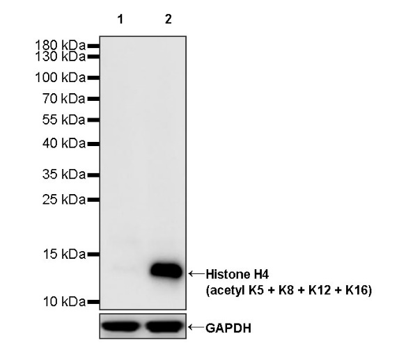
WB result of Histone H4 (acetyl K5 + K8 + K12 + K16) Recombinant Rabbit mAb
Primary antibody: Histone H4 (acetyl K5 + K8 + K12 + K16) Recombinant Rabbit mAb at 1/1000 dilution
Lane 1: untreated NIH/3T3 whole cell lysate 20 µg
Lane 2: NIH/3T3 treated with 500 ng/ml TSA for 4 hours whole cell lysate 20 µg
Secondary antibody: Goat Anti-rabbit IgG, (H+L), HRP conjugated at 1/10000 dilution
Predicted MW: 11 kDa
Observed MW: 13 kDa
Dot blot result of Histone H4 (acetyl K5 + K8 + K12 + K16) Recombinant Rabbit mAb
Lane 1: Histone H4 (acetyl K5 + K8 + K12 + K16) peptide
Lane 2: Histone H4 (acetyl K5) peptide
Lane 3: Histone H4 (acetyl K8) peptide
Lane 4: Histone H4 (acetyl K12) peptide
Lane 5: Histone H4 (acetyl K16) peptide
Lane 6: Histone H4 unmodified peptide
Primary antibody: Histone H4 (acetyl K5 + K8 + K12 + K16) Recombinant Rabbit mAb at 1/1000 dilution
Secondary antibody: Goat Anti-rabbit IgG, (H+L), HRP conjugated at 1/10000 dilution
IHC shows positive staining in paraffin-embedded human colon. Anti-Histone H4 (acetyl K5 + K8 + K12 + K16) antibody was used at 1/200 dilution, followed by a HRP Polymer for Mouse & Rabbit IgG (ready to use). Counterstained with hematoxylin. Heat mediated antigen retrieval with Tris/EDTA buffer pH9.0 was performed before commencing with IHC staining protocol.
IHC shows positive staining in paraffin-embedded human kidney. Anti-Histone H4 (acetyl K5 + K8 + K12 + K16) antibody was used at 1/200 dilution, followed by a HRP Polymer for Mouse & Rabbit IgG (ready to use). Counterstained with hematoxylin. Heat mediated antigen retrieval with Tris/EDTA buffer pH9.0 was performed before commencing with IHC staining protocol.
IHC shows positive staining in paraffin-embedded human breast cancer. Anti-Histone H4 (acetyl K5 + K8 + K12 + K16) antibody was used at 1/200 dilution, followed by a HRP Polymer for Mouse & Rabbit IgG (ready to use). Counterstained with hematoxylin. Heat mediated antigen retrieval with Tris/EDTA buffer pH9.0 was performed before commencing with IHC staining protocol.
IHC shows positive staining in paraffin-embedded human endometrial cancer. Anti-Histone H4 (acetyl K5 + K8 + K12 + K16) antibody was used at 1/200 dilution, followed by a HRP Polymer for Mouse & Rabbit IgG (ready to use). Counterstained with hematoxylin. Heat mediated antigen retrieval with Tris/EDTA buffer pH9.0 was performed before commencing with IHC staining protocol.
IHC shows positive staining in paraffin-embedded mouse stomach. Anti-Histone H4 (acetyl K5 + K8 + K12 + K16) antibody was used at 1/200 dilution, followed by a HRP Polymer for Mouse & Rabbit IgG (ready to use). Counterstained with hematoxylin. Heat mediated antigen retrieval with Tris/EDTA buffer pH9.0 was performed before commencing with IHC staining protocol.
IHC shows positive staining in paraffin-embedded rat stomach. Anti-Histone H4 (acetyl K5 + K8 + K12 + K16) antibody was used at 1/200 dilution, followed by a HRP Polymer for Mouse & Rabbit IgG (ready to use). Counterstained with hematoxylin. Heat mediated antigen retrieval with Tris/EDTA buffer pH9.0 was performed before commencing with IHC staining protocol.
ICC analysis of HeLa cells treated with TSA (500ng/ml,4h) (top panel) and HeLa cells untreated with TSA (500ng/ml,4h) (below panel). Anti- Histone H4 (acetyl K5 + K8 + K12 + K16) antibody was used at 1/500 dilution (Green) and incubated overnight at 4°C. Goat polyclonal Antibody to Rabbit IgG - H&L (Alexa Fluor® 488) was used as secondary antibody at 1/1000 dilution. The cells were fixed with 100% ice-cold methanol and permeabilized with 0.1% PBS-Triton X-100. Nuclei were counterstained with DAPI (Blue). Counterstain with tubulin (Red).
Chromatin immunoprecipitation (ChIP) was
performed on HeLa+TSA (500ng/ml,4h) (+) cells
cross - linked with 1% formaldehyde for 10 min,
then chromatin was fragmented by sonication.
Parallel reactions used Histone H4 (acetyl K5 +
K8 + K12 + K16) Recombinant Rabbit mAb (S-902-37)
and Rabbit mAb IgG Isotype Control (SDT-R173)
at 1:50 for immunoprecipitation.
Post - immunoprecipitation, both samples
were washed, eluted, and cross - links
reversed. Purified DNA was analyzed by qPCR.
qPCR (%input: immunoprecipitated DNA/input DNA)
showed the enrichment of RPL30, GAPDH, MYOD1,
AFM, SAT-α and SAT-2 in Histone H4 (acetyl K5 + K8 +
K12 + K16) Recombinant Rabbit mAb (S-902-37)
-immunoprecipitated sample.
 您现在的位置: