
PBS, 40% Glycerol, 0.05% BSA, 0.03% Proclin 300
12 months from date of receipt / reconstitution, -20 °C as supplied
| 应用 | 稀释度 |
|---|---|
| WB | 1:5000 |
| ICC | 1:500-1:2000 |
| ICFCM | 1:5000 |
A polyhistidine-tag is an amino acid motif in proteins that typically consists of at least six histidine (His) residues, often at the N- or C-terminus of the protein. The total number of histidine residues may vary in the tag from as low as two, to as high as 10 or more His residues.
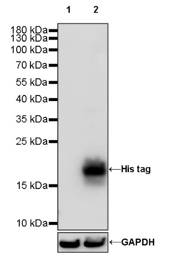
WB result of His tag Rabbit pAb
Primary antibody: His tag Rabbit pAb at 1/5000 dilution
Lane 1: untreated 293T whole cell lysate 20 µg
Lane 2: 293T transfected with myc-his tag whole cell lysate 20 µg
Secondary antibody: Goat Anti-rabbit IgG, (H+L), HRP conjugated at 1/10000 dilution
Predicted MW: 24 kDa
Observed MW: 20 kDa
WB result of His tag Rabbit pAb
Primary antibody: His tag Rabbit pAb at 1/5000 dilution
Lane 1: Angiopoietin-1 (277-498) His Tag Protein, Mouse 10 ng
Secondary antibody: Goat Anti-rabbit IgG, (H+L), HRP conjugated at 1/10000 dilution
Predicted MW: 28~33 kDa
Observed MW: 28 kDa
Flow cytometric analysis of 4% PFA fixed 90% methanol permeabilized Myc, His transfected 293T (Human embryonic kidney epithelial cell) labelling His tag antibody at 1/5000 (0.01 μg) dilution/ (right panel) compared with a Rabbit IgG Isotype Control / (left panel). Goat Anti-Rabbit IgG Alexa Fluor® 488 was used as the secondary antibody.
ICC shows positive staining in Histone H3-His Tag transfected 293T cells (top panel) and negative staining in vector-transfected 293T cells (below panel). Anti-His tag Rabbit Polyclonal antibody was used at 1/500 dilution (Green) and incubated overnight at 4°C. Goat polyclonal Antibody to Rabbit IgG - H&L (Alexa Fluor® 488) was used as secondary antibody at 1/1000 dilution. The cells were fixed with 4% PFA and permeabilized with 0.1% PBS-Triton X-100. Nuclei were counterstained with DAPI (Blue). Counterstain with tubulin (Red).
 您现在的位置: