
12 months from date of receipt / reconstitution, -20 °C as supplied
| 应用 | 稀释度 |
|---|---|
| WB | 1:4000 |
| IHC-P | 1:500 |
| ICC | 1:2000 |
| ICFCM | 1:200 |
The Helios protein belongs to the Ikaros family of zinc finger proteins, which are hematopoietic-specific transcription factors involved in the regulation of lymphocyte development. The Helios protein serves as an epigenetic regulator, selectively expressing some genes while suppressing others. It influences gene expression by modulating chromatin structure, thereby controlling DNA accessibility. This regulatory mechanism is vital for normal cell growth and differentiation. The Helios protein exhibits a unique dual role in leukemia. Its deficiency has been linked to the development of acute B-lymphoblastic leukemia (B-ALL), while its overexpression promotes acute myeloid leukemia (AML).
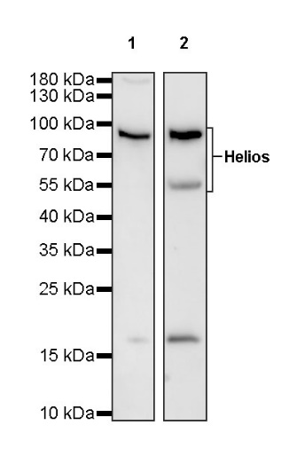
WB result of Helios Recombinant Rabbit mAb
Primary antibody: Helios Recombinant Rabbit mAb at 1/4000 dilution
Lane 1: Jurkat whole cell lysate 20 µg
Lane 2: Raji whole cell lysate 20 µg
Secondary antibody: Goat Anti-rabbit IgG, (H+L), HRP conjugated at 1/10000 dilution
Predicted MW: 60 kDa
Observed MW: 55, 85 kDa
WB result of Helios Recombinant Rabbit mAb
Primary antibody: Helios Recombinant Rabbit mAb at 1/4000 dilution
Lane 1: EL4 whole cell lysate 20 µg
Lane 2: mouse thymus lysate 20 µg
Lane 3: mouse spleen lysate 20 µg
Secondary antibody: Goat Anti-rabbit IgG, (H+L), HRP conjugated at 1/10000 dilution
Predicted MW: 60 kDa
Observed MW: 55, 63~70, 85 kDa
WB result of Helios Recombinant Rabbit mAb
Primary antibody: Helios Recombinant Rabbit mAb at 1/4000 dilution
Lane 1: rat thymus lysate 20 µg
Lane 3: rat spleen lysate 20 µg
Secondary antibody: Goat Anti-rabbit IgG, (H+L), HRP conjugated at 1/10000 dilution
Predicted MW: 60 kDa
Observed MW: 55, 85 kDa
Flow cytometric analysis of 4% PFA fixed 90% methanol permeabilized Jurkat (Human T cell leukemia T lymphocyte) labelling Helios antibody at 1/200 dilution (1 μg)/ (Red) compared with a Rabbit monoclonal IgG (Black) isotype control and an unlabelled control (cells without incubation with primary antibody and secondary antibody) (Blue). Goat Anti - Rabbit IgG Alexa Fluor® 488 was used as the secondary antibody.
IHC shows positive staining in paraffin-embedded human thymus. Anti-Helios antibody was used at 1/500 dilution, followed by a HRP Polymer for Mouse & Rabbit IgG (ready to use). Counterstained with hematoxylin. Heat mediated antigen retrieval with Tris/EDTA buffer pH9.0 was performed before commencing with IHC staining protocol.
IHC shows positive staining in paraffin-embedded human cervical squamous cell carcinoma. Anti-Helios antibody was used at 1/500 dilution, followed by a HRP Polymer for Mouse & Rabbit IgG (ready to use). Counterstained with hematoxylin. Heat mediated antigen retrieval with Tris/EDTA buffer pH9.0 was performed before commencing with IHC staining protocol.
IHC shows positive staining in paraffin-embedded human endometrial cancer. Anti-Helios antibody was used at 1/500 dilution, followed by a HRP Polymer for Mouse & Rabbit IgG (ready to use). Counterstained with hematoxylin. Heat mediated antigen retrieval with Tris/EDTA buffer pH9.0 was performed before commencing with IHC staining protocol.
IHC shows positive staining in paraffin-embedded human thyroid carcinoma. Anti-Helios antibody was used at 1/500 dilution, followed by a HRP Polymer for Mouse & Rabbit IgG (ready to use). Counterstained with hematoxylin. Heat mediated antigen retrieval with Tris/EDTA buffer pH9.0 was performed before commencing with IHC staining protocol.
ICC shows positive staining in Jurkat cells. Anti- Helios antibody was used at 1/2000 dilution (Green) and incubated overnight at 4°C. Goat polyclonal Antibody to Rabbit IgG - H&L (Alexa Fluor® 488) was used as secondary antibody at 1/1000 dilution. The cells were fixed with 4% PFA and permeabilized with 0.1% PBS-Triton X-100. Nuclei were counterstained with DAPI (Blue). Counterstain with tubulin (Red).
 您现在的位置: