
PBS, 40% Glycerol, 0.05% BSA, 0.03% Proclin 300
12 months from date of receipt / reconstitution, -20 °C as supplied
| 应用 | 稀释度 |
|---|---|
| WB | 1:1000 |
| IP | 1:200 |
| IHC-P | 1:1000-1:2000 |
HLA-A, short for Human Leukocyte Antigen-A, is a crucial component of the Human Major Histocompatibility Complex (MHC) Class I molecules. HLA-A molecules play a central role in the immune system by presenting peptides derived from the endoplasmic reticulum lumen to T-cell receptors, triggering immune responses. HLA-A is critical in determining tissue compatibility between individuals. In organ and bone marrow transplantations, the matching degree of HLA-A is a crucial factor in assessing the risk of transplant rejection. Certain alleles of HLA-A have been associated with the risk of developing specific diseases, such as ankylosing spondylitis and other rheumatic diseases. In transplantation procedures involving bone marrow, kidneys, etc., HLA-A typing is a crucial step to ensure transplant success and minimize rejection risks.
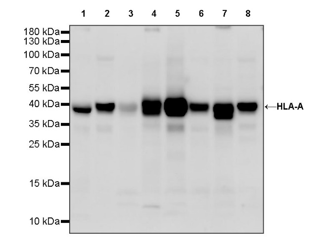
WB result of HLA-A Rabbit pAb
Primary antibody: HLA-A Rabbit pAb at 1/1000 dilution
Lane 1: THP-1 whole cell lysate 20 µg
Lane 2: Raji whole cell lysate 20 µg
Lane 3: A549 whole cell lysate 20 µg
Lane 4: HL-60 whole cell lysate 20 µg
Lane 5: A431 whole cell lysate 20 µg
Lane 6: Jurkat whole cell lysate 20 µg
Lane 7: HepG2 whole cell lysate 20 µg
Lane 8: Ramos whole cell lysate 20 µg
Secondary antibody: Goat Anti-rabbit IgG, (H+L), HRP conjugated at 1/10000 dilution
Predicted MW: 41 kDa
Observed MW: 40 kDa
HLA-A Rabbit pAb at 1/200 dilution (1 µg) immunoprecipitating HLA-A in 0.4 mg Ramos whole cell lysate.
Western blot was performed on the immunoprecipitate using HLA-A Rabbit pAb at 1/1000 dilution.
Secondary antibody (HRP) for IP was used at 1/1000 dilution.
Lane 1: Ramos whole cell lysate 5 µg (Input)
Lane 2: HLA-A Rabbit pAb IP in Ramos whole cell lysate
Lane 3: Rabbit monoclonal IgG IP in Ramos whole cell lysate
Predicted MW: 41 kDa
Observed MW: 40 kDa
IHC shows positive staining in paraffin-embedded human liver. Anti- HLA-A antibody was used at 1/1000 dilution, followed by a HRP Polymer for Mouse & Rabbit IgG (ready to use). Counterstained with hematoxylin. Heat mediated antigen retrieval with Tris/EDTA buffer pH9.0 was performed before commencing with IHC staining protocol.
IHC shows positive staining in paraffin-embedded human tonsil. Anti- HLA-A antibody was used at 1/1000 dilution, followed by a HRP Polymer for Mouse & Rabbit IgG (ready to use). Counterstained with hematoxylin. Heat mediated antigen retrieval with Tris/EDTA buffer pH9.0 was performed before commencing with IHC staining protocol.
IHC shows positive staining in paraffin-embedded human hepatocellular carcinoma. Anti- HLA-A antibody was used at 1/1000 dilution, followed by a HRP Polymer for Mouse & Rabbit IgG (ready to use). Counterstained with hematoxylin. Heat mediated antigen retrieval with Tris/EDTA buffer pH9.0 was performed before commencing with IHC staining protocol.
IHC shows positive staining in paraffin-embedded human bladder cancer. Anti- HLA-A antibody was used at 1/1000 dilution, followed by a HRP Polymer for Mouse & Rabbit IgG (ready to use). Counterstained with hematoxylin. Heat mediated antigen retrieval with Tris/EDTA buffer pH9.0 was performed before commencing with IHC staining protocol.
IHC shows positive staining in paraffin-embedded human endometrial cancer. Anti- HLA-A antibody was used at 1/1000 dilution, followed by a HRP Polymer for Mouse & Rabbit IgG (ready to use). Counterstained with hematoxylin. Heat mediated antigen retrieval with Tris/EDTA buffer pH9.0 was performed before commencing with IHC staining protocol.
 您现在的位置: