
12 months from date of receipt / reconstitution, -20 °C as supplied
| 应用 | 稀释度 |
|---|---|
| WB | 1:1000 |
| IHC-P | 1:1000 |
| ICC | 1:500 |
| ICFCM | 1:50 |
C3 plays a pivotal role in initiating both the classical and alternative pathways of the complement system. Upon activation by C3 convertases, C3 is cleaved into C3a and C3b fragments. C3b covalently binds to cell surface carbohydrates or immune aggregates, while C3a acts as an anaphylatoxin mediating local inflammatory responses. Beyond inflammation, C3 regulates the intensity and direction of immune responses through interactions with other complement components. Detection of C3 levels is valuable for diagnosing, monitoring, and assessing prognosis in various diseases, such as autoimmune disorders, infectious diseases, and cardiovascular conditions. As a crucial component of the complement system, C3 is a significant target for drug development and immunotherapy research.
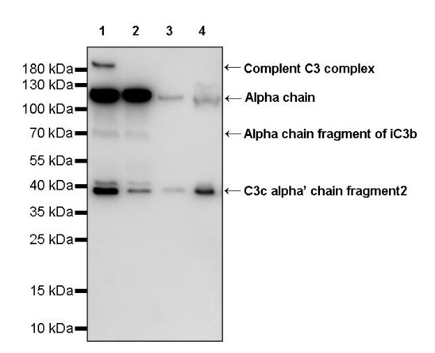
WB result of C3c Recombinant Rabbit mAb
Primary antibody: C3c Recombinant Rabbit mAb at 1/1000 dilution
Lane 1: human serum lysate 5 µg
Lane 2: human plasma lysate 5 µg
Lane 3: mouse plasma lysate 20 µg
Lane 4: rat serum lysate 20 µg
Secondary antibody: Goat Anti-rabbit IgG, (H+L), HRP conjugated at 1/10000 dilution
Predicted MW: 187 kDa
Observed MW: 38, 70, 115, 187 kDa
Flow cytometric analysis of 4% PFA fixed 90% methanol permeabilized HepG2 (Human hepatocellular carcinoma epithelial cell) labelling C3c antibody at 1/50 dilution (1 μg)/ (Red) compared with a Rabbit monoclonal IgG (Black) isotype control and an unlabelled control (cells without incubation with primary antibody and secondary antibody) (Blue). Goat Anti - Rabbit IgG Alexa Fluor® 488 was used as the secondary antibody.
IHC shows positive staining in paraffin-embedded human kidney. Anti-C3c antibody was used at 1/1000 dilution, followed by a HRP Polymer for Mouse & Rabbit IgG (ready to use). Counterstained with hematoxylin. Heat mediated antigen retrieval with Tris/EDTA buffer pH9.0 was performed before commencing with IHC staining protocol.
IHC shows positive staining in paraffin-embedded human liver. Anti-C3c antibody was used at 1/1000 dilution, followed by a HRP Polymer for Mouse & Rabbit IgG (ready to use). Counterstained with hematoxylin. Heat mediated antigen retrieval with Tris/EDTA buffer pH9.0 was performed before commencing with IHC staining protocol.
IHC shows positive staining in paraffin-embedded human lung. Anti-C3c antibody was used at 1/1000 dilution, followed by a HRP Polymer for Mouse & Rabbit IgG (ready to use). Counterstained with hematoxylin. Heat mediated antigen retrieval with Tris/EDTA buffer pH9.0 was performed before commencing with IHC staining protocol.
IHC shows positive staining in paraffin-embedded mouse kidney. Anti-C3c antibody was used at 1/1000 dilution, followed by a HRP Polymer for Mouse & Rabbit IgG (ready to use). Counterstained with hematoxylin. Heat mediated antigen retrieval with Tris/EDTA buffer pH9.0 was performed before commencing with IHC staining protocol.
IHC shows positive staining in paraffin-embedded rat testis. Anti-C3c antibody was used at 1/1000 dilution, followed by a HRP Polymer for Mouse & Rabbit IgG (ready to use). Counterstained with hematoxylin. Heat mediated antigen retrieval with Tris/EDTA buffer pH9.0 was performed before commencing with IHC staining protocol.
ICC shows positive staining in HepG2 cells. Anti-C3c antibody was used at 1/500 dilution (Green) and incubated overnight at 4°C. Goat polyclonal Antibody to Rabbit IgG - H&L (Alexa Fluor® 488) was used as secondary antibody at 1/1000 dilution. The cells were fixed with 100% ice-cold methanol and permeabilized with 0.1% PBS-Triton X-100. Nuclei were counterstained with DAPI (Blue). Counterstain with tubulin (Red).
 您现在的位置: