
PBS, 40% Glycerol, 0.05% BSA, 0.03% Proclin 300
12 months from date of receipt / reconstitution, -20 °C as supplied
| 应用 | 稀释度 |
|---|---|
| WB | 1:1000 |
| IP | 1:50 |
| IHC-P | 1:250 |
| ICC | 1:500 |
| ICFCM | 1:500 |
Smad1, or Smad Homolog 1, is a vital protein involved in cellular signal transduction, particularly within the TGF-β (Transforming Growth Factor-β) signaling pathway. Smad1 serves as a key transcription factor in the TGF-β signaling pathway, facilitating the transmission of extracellular signals from the cell membrane to the nucleus, where it regulates the transcription of target genes. Research indicates that Smad1 plays a crucial role in bone development and metabolism. For instance, BMP9 activates the Smad1 signaling pathway, inhibiting osteoblast senescence and thereby reducing age-related bone loss in mice. The role of Smad1 in tumorigenesis is also gaining attention. Studies have found that Atm-mediated phosphorylation of Smad1 is important in the DNA damage response and tumorigenesis. In ophthalmology research, Smad1 has been shown to have a protective role in mouse refractive development and the formation of form-deprivation myopia.
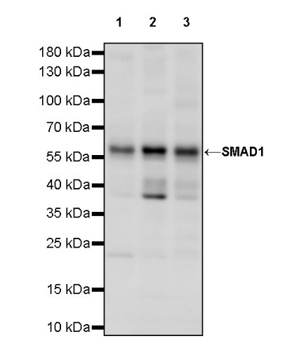
WB result of SMAD1 Rabbit pAb
Primary antibody: SMAD1 Rabbit pAb at 1/1000 dilution
Lane 1: HeLa whole cell lysate 20 µg
Lane 2: HEK-293 whole cell lysate 20 µg
Lane 3: HT-1080 whole cell lysate 20 µg
Secondary antibody: Goat Anti-rabbit IgG, (H+L), HRP conjugated at 1/10000 dilution
Predicted MW: 52 kDa
Observed MW: 60 kDa
WB result of SMAD1 Rabbit pAb
Primary antibody: SMAD1 Rabbit pAb at 1/1000 dilution
Lane 1: NIH/3T3 whole cell lysate 20 µg
Secondary antibody: Goat Anti-rabbit IgG, (H+L), HRP conjugated at 1/10000 dilution
Predicted MW: 52 kDa
Observed MW: 60 kDa
Flow cytometric analysis of 4% PFA fixed 90% methanol permeabilized HeLa (Human cervix adenocarcinoma epithelial cell) labelling SMAD1 antibody at 1/500 dilution (0.1 μg)/ (Red) compared with a Rabbit monoclonal IgG (Black) isotype control and an unlabelled control (cells without incubation with primary antibody and secondary antibody) (Blue). Goat Anti - Rabbit IgG Alexa Fluor® 488 was used as the secondary antibody.
SMAD1 Rabbit pAb at 1/50 dilution (1 µg) immunoprecipitating SMAD1 in 0.4 mg HeLa whole cell lysate.
Western blot was performed on the immunoprecipitate using SMAD1 Rabbit pAb at 1/1000 dilution.
Secondary antibody (HRP) for IP was used at 1/1000 dilution.
Lane 1: HeLa whole cell lysate 40 µg (Input)
Lane 2: SMAD1 Rabbit pAb IP in HeLa whole cell lysate
Lane 3: Rabbit monoclonal IgG IP in HeLa whole cell lysate
Predicted MW: 52 kDa
Observed MW: 60 kDa
IHC shows positive staining in paraffin-embedded human tonsil. Anti-SMAD1 antibody was used at 1/250 dilution, followed by a HRP Polymer for Mouse & Rabbit IgG (ready to use). Counterstained with hematoxylin. Heat mediated antigen retrieval with Tris/EDTA buffer pH9.0 was performed before commencing with IHC staining protocol.
IHC shows positive staining in paraffin-embedded human stomach. Anti-SMAD1 antibody was used at 1/250 dilution, followed by a HRP Polymer for Mouse & Rabbit IgG (ready to use). Counterstained with hematoxylin. Heat mediated antigen retrieval with Tris/EDTA buffer pH9.0 was performed before commencing with IHC staining protocol.
IHC shows positive staining in paraffin-embedded human ovarian cancer. Anti-SMAD1 antibody was used at 1/250 dilution, followed by a HRP Polymer for Mouse & Rabbit IgG (ready to use). Counterstained with hematoxylin. Heat mediated antigen retrieval with Tris/EDTA buffer pH9.0 was performed before commencing with IHC staining protocol.
IHC shows positive staining in paraffin-embedded rat colon. Anti-SMAD1 antibody was used at 1/250 dilution, followed by a HRP Polymer for Mouse & Rabbit IgG (ready to use). Counterstained with hematoxylin. Heat mediated antigen retrieval with Tris/EDTA buffer pH9.0 was performed before commencing with IHC staining protocol.
ICC shows positive staining in HeLa cells. Anti-SMAD1 antibody was used at 1/500 dilution (Green) and incubated overnight at 4°C. Goat polyclonal Antibody to Rabbit IgG - H&L (Alexa Fluor® 488) was used as secondary antibody at 1/1000 dilution. The cells were fixed with 4% PFA and permeabilized with 0.1% PBS-Triton X-100. Nuclei were counterstained with DAPI (Blue). Counterstain with tubulin (Red).
 您现在的位置: