
12 months from date of receipt / reconstitution, -20 °C as supplied
| 应用 | 稀释度 |
|---|---|
| Dot Blot | 1:1000 |
| WB | 1:1000 |
| ICC | 1:500 |
| ICFCM | 1:500 |
H3K9me1 refers to the monomethylation of the ninth lysine residue on histone H3. This modification can either activate or repress gene expression, depending on the cellular context, developmental stage, and environmental cues. In some instances, H3K9me1 may contribute to maintaining an open chromatin state, facilitating the binding of transcription factors to DNA and thereby activating gene transcription. Alternatively, it can associate with repressive complexes, leading to chromatin condensation and gene repression. The levels of H3K9me1 are tightly regulated by a balance of enzymatic activities, including methyltransferases (such as SETDB1 and G9a) that add methyl groups and demethylases (like LSD1) that remove them. These enzymes precisely control the extent of H3K9me1 modification, thereby modulating gene expression and cellular behaviors.
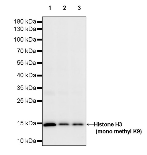
WB result of Histone H3 (mono methyl K9) Recombinant Rabbit mAb
Primary antibody: Histone H3 (mono methyl K9) Recombinant Rabbit mAb at 1/1000 dilution
Lane 1: HeLa whole cell lysate 20 µg
Lane 2: MCF7 whole cell lysate 20 µg
Lane 3: Jurkat whole cell lysate 20 µg
Secondary antibody: Goat Anti-rabbit IgG, (H+L), HRP conjugated at 1/10000 dilution
Predicted MW: 15 kDa
Observed MW: 15 kDa
WB result of Histone H3 (mono methyl K9) Recombinant Rabbit mAb
Primary antibody: Histone H3 (mono methyl K9) Recombinant Rabbit mAb at 1/1000 dilution
Lane 1: Neuro-2a whole cell lysate 20 µg
Lane 2: NIH/3T3 whole cell lysate 20 µg
Lane 3: C2C12 whole cell lysate 20 µg
Secondary antibody: Goat Anti-rabbit IgG, (H+L), HRP conjugated at 1/10000 dilution
Predicted MW: 15 kDa
Observed MW: 15 kDa
WB result of Histone H3 (mono methyl K9) Recombinant Rabbit mAb
Primary antibody: Histone H3 (mono methyl K9) Recombinant Rabbit mAb at 1/1000 dilution
Lane 1: C6 whole cell lysate 20 µg
Secondary antibody: Goat Anti-rabbit IgG, (H+L), HRP conjugated at 1/10000 dilution
Predicted MW: 15 kDa
Observed MW: 15 kDa
Flow cytometric analysis of 4% PFA fixed 90% methanol permeabilized HeLa (Human cervix adenocarcinoma epithelial cell) labelling Histone H3 (mono methyl K9) antibody at 1/500 dilution (0.1 μg)/ (Red) compared with a Rabbit monoclonal IgG (Black) isotype control and an unlabelled control (cells without incubation with primary antibody and secondary antibody) (Blue). Goat Anti - Rabbit IgG Alexa Fluor® 488 was used as the secondary antibody.
Dot blot result of Histone H3 (mono methyl K9) Recombinant Rabbit mAb
Lane 1: H3K9me1 peptide
Lane 2: H3K9me2 peptide
Lane 3: H3K9me3 peptide
Lane 4: H3K4me1 peptide
Lane 5: H3K9un peptide
Primary antibody: Histone H3 (mono methyl K9) Recombinant Rabbit mAb at 1/1000 dilution
Secondary antibody: Goat Anti-rabbit IgG, (H+L), HRP conjugated at 1/10000 dilution
ICC shows positive staining in HeLa cells. Anti- Histone H3 (mono methyl K9) antibody was used at 1/500 dilution (Green) and incubated overnight at 4°C. Goat polyclonal Antibody to Rabbit IgG - H&L (Alexa Fluor® 488) was used as secondary antibody at 1/1000 dilution. The cells were fixed with 100% ice-cold methanol and permeabilized with 0.1% PBS-Triton X-100. Nuclei were counterstained with DAPI (Blue). Counterstain with tubulin (Red).
 您现在的位置: