
12 months from date of receipt / reconstitution, -20 °C as supplied
| 应用 | 稀释度 |
|---|---|
| WB | 1:1000 |
| IHC-P | 1:500 |
| IF | 1:500 |
Myelin oligodendrocyte glycoprotein (MOG) is a glycoprotein believed to be important in the myelination of nerves in the central nervous system (CNS). It is speculated to serve as a necessary "adhesion molecule" to provide structural integrity to the myelin sheath and is known to develop late on the oligodendrocyte. Interest in MOG has centered on its role in demyelinating diseases. Some of them are not-inflammatory, such as adrenoleukodystrophy, vanishing white matter disease, and Rubella induced mental retardation.
WB result of Myelin Oligodendrocyte Glycoprotein (MOG) Recombinant Rabbit mAb
Primary antibody: Myelin Oligodendrocyte Glycoprotein (MOG) Recombinant Rabbit mAb at 1/1000 dilution
Lane 1: mouse lung lysate 20 µg
Lane 2: mouse brain lysate 20 µg
Negative control: mouse lung lysate
Secondary antibody: Goat Anti-rabbit IgG, (H+L), HRP conjugated at 1/10000 dilution
Predicted MW: 28 kDa
Observed MW: 25 kDa
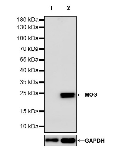
WB result of Myelin Oligodendrocyte Glycoprotein (MOG) Recombinant Rabbit mAb
Primary antibody: Myelin Oligodendrocyte Glycoprotein (MOG) Recombinant Rabbit mAb at 1/1000 dilution
Lane 1: rat brain lysate 20 µg
Secondary antibody: Goat Anti-rabbit IgG, (H+L), HRP conjugated at 1/10000 dilution
Predicted MW: 28 kDa
Observed MW: 25 kDa
IHC shows positive staining in paraffin-embedded human cerebral cortex. Anti-Myelin Oligodendrocyte Glycoprotein (MOG) antibody was used at 1/500 dilution, followed by a HRP Polymer for Mouse & Rabbit IgG (ready to use). Counterstained with hematoxylin. Heat mediated antigen retrieval with Tris/EDTA buffer pH9.0 was performed before commencing with IHC staining protocol.
Negative control: IHC shows negative staining in paraffin-embedded human colon. Anti-Myelin Oligodendrocyte Glycoprotein (MOG) antibody was used at 1/500 dilution, followed by a HRP Polymer for Mouse & Rabbit IgG (ready to use). Counterstained with hematoxylin. Heat mediated antigen retrieval with Tris/EDTA buffer pH9.0 was performed before commencing with IHC staining protocol.
Negative control: IHC shows negative staining in paraffin-embedded human colon cancer. Anti-Myelin Oligodendrocyte Glycoprotein (MOG) antibody was used at 1/500 dilution, followed by a HRP Polymer for Mouse & Rabbit IgG (ready to use). Counterstained with hematoxylin. Heat mediated antigen retrieval with Tris/EDTA buffer pH9.0 was performed before commencing with IHC staining protocol.
IHC shows positive staining in paraffin-embedded mouse cerebral cortex. Anti-Myelin Oligodendrocyte Glycoprotein (MOG) antibody was used at 1/500 dilution, followed by a HRP Polymer for Mouse & Rabbit IgG (ready to use). Counterstained with hematoxylin. Heat mediated antigen retrieval with Tris/EDTA buffer pH9.0 was performed before commencing with IHC staining protocol.
IHC shows positive staining in paraffin-embedded rat cerebral cortex. Anti-Myelin Oligodendrocyte Glycoprotein (MOG) antibody was used at 1/500 dilution, followed by a HRP Polymer for Mouse & Rabbit IgG (ready to use). Counterstained with hematoxylin. Heat mediated antigen retrieval with Tris/EDTA buffer pH9.0 was performed before commencing with IHC staining protocol.
IF shows positive staining in paraffin-embedded human cerebral cortex. Anti- Myelin Oligodendrocyte Glycoprotein antibody was used at 1/500 dilution (Green) and incubated overnight at 4°C. Goat polyclonal Antibody to Rabbit IgG - H&L (Alexa Fluor® 488) was used as secondary antibody at 1/1000 dilution. Counterstained with DAPI (Blue). Heat mediated antigen retrieval with EDTA buffer pH9.0 was performed before commencing with IF staining protocol.
IF shows positive staining in paraffin-embedded mouse cerebral cortex. Anti- Myelin Oligodendrocyte Glycoprotein antibody was used at 1/500 dilution (Green) and incubated overnight at 4°C. Goat polyclonal Antibody to Rabbit IgG - H&L (Alexa Fluor® 488) was used as secondary antibody at 1/1000 dilution. Counterstained with DAPI (Blue). Heat mediated antigen retrieval with EDTA buffer pH9.0 was performed before commencing with IF staining protocol.
 您现在的位置: