
12 months from date of receipt / reconstitution, -20 °C as supplied
| 应用 | 稀释度 |
|---|---|
| WB | 1:1000 |
| IHC-P | 1:100-1:200 |
| IP | 1:50 |
The NDUFS8 protein is a subunit of NADH dehydrogenase (ubiquinone) also known as Complex I, which is located in the mitochondrial inner membrane and is the largest of the five complexes of the electron transport chain. Mutations in NDUFS8 have been associated with mitochondrial diseases, which can cause any one of a clinically heterogeneous group of disorders arising from dysfunction of the mitochondrial respiratory chain. The phenotypic spectrum ranges from isolated diseases affecting single organs to severe multisystem disorders. NDUFS8 mutations have also been associated with Leigh syndrome.
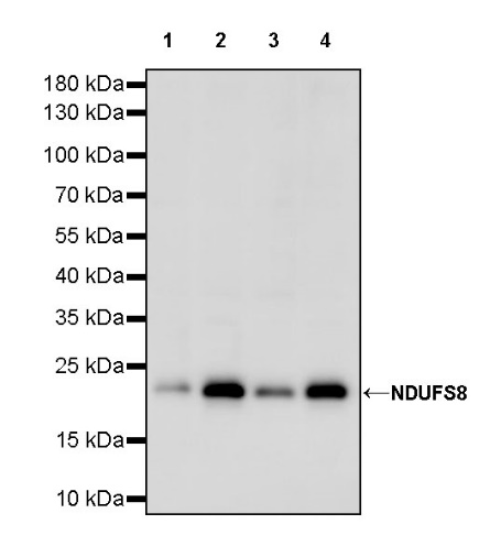
WB result of NDUFS8 Recombinant Rabbit mAb
Primary antibody: NDUFS8 Recombinant Rabbit mAb at 1/1000 dilution
Lane 1: HeLa whole cell lysate 20 µg
Lane 2: HepG2 whole cell lysate 20 µg
Lane 3: Jurkat whole cell lysate 20 µg
Lane 4: 293T whole cell lysate 20 µg
Secondary antibody: Goat Anti-rabbit IgG, (H+L), HRP conjugated at 1/10000 dilution
Predicted MW: 23 kDa
Observed MW: 23 kDa
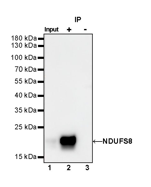
NDUFS8 Rabbit mAb at 1/50 dilution (1 µg) immunoprecipitating NDUFS8 in 0.4 mg HepG2 whole cell lysate.
Western blot was performed on the immunoprecipitate using NDUFS8 Rabbit mAb at 1/1000 dilution.
Secondary antibody (HRP) for IP was used at 1/1000 dilution.
Lane 1: HepG2 whole cell lysate 20 µg (Input)
Lane 2: NDUFS8 Rabbit mAb IP in HepG2 whole cell lysate
Lane 3: Rabbit monoclonal IgG IP in HepG2 whole cell lysate
Predicted MW: 23 kDa
Observed MW: 23 kDa
IHC shows positive staining in paraffin-embedded human cerebral cortex. Anti-NDUFS8 antibody was used at 1/200 dilution, followed by a HRP Polymer for Mouse & Rabbit IgG (ready to use). Counterstained with hematoxylin. Heat mediated antigen retrieval with Tris/EDTA buffer pH9.0 was performed before commencing with IHC staining protocol.
IHC shows positive staining in paraffin-embedded human cardiac muscle. Anti-NDUFS8 antibody was used at 1/100 dilution, followed by a HRP Polymer for Mouse & Rabbit IgG (ready to use). Counterstained with hematoxylin. Heat mediated antigen retrieval with Tris/EDTA buffer pH9.0 was performed before commencing with IHC staining protocol.
IHC shows positive staining in paraffin-embedded human kidney. Anti-NDUFS8 antibody was used at 1/100 dilution, followed by a HRP Polymer for Mouse & Rabbit IgG (ready to use). Counterstained with hematoxylin. Heat mediated antigen retrieval with Tris/EDTA buffer pH9.0 was performed before commencing with IHC staining protocol.
IHC shows positive staining in paraffin-embedded human colon cancer. Anti-NDUFS8 antibody was used at 1/100 dilution, followed by a HRP Polymer for Mouse & Rabbit IgG (ready to use). Counterstained with hematoxylin. Heat mediated antigen retrieval with Tris/EDTA buffer pH9.0 was performed before commencing with IHC staining protocol.
 您现在的位置: