
12 months from date of receipt / reconstitution, -20 °C as supplied
| 应用 | 稀释度 |
|---|---|
| WB | 1:5000 |
| IHC-P | 1:500 |
| FCM | 1:50 |
ICAM-1, also known as Intercellular Cell Adhesion Molecule-1 and CD54, is a vital member of the immunoglobulin superfamily (IGSF) of adhesion molecules. It exists in two forms: soluble (sICAM-1) and membrane-bound (mICAM-1). sICAM-1 is derived from the proteolytic cleavage of mICAM-1 and released into the bloodstream, reflecting local ICAM-1 expression levels. ICAM-1 facilitates adhesion between leukocytes and endothelial cells by binding to specific receptors such as LFA-1 and Mac-1, enabling cell migration. ICAM-1 participates in cell signaling, activation, growth, differentiation, immune responses, inflammation, angiogenesis, and tumor metastasis. During inflammation, ICAM-1 upregulation enhances the immune system's ability to eliminate foreign antigens and tumor cells. ICAM-1 is intimately linked to the development of various diseases, including atherosclerosis, rheumatoid arthritis, multiple sclerosis, inflammatory bowel diseases, and multiple cancers (e.g., multiple myeloma, triple-negative breast cancer, thyroid cancer). ICAM-1 upregulation promotes leukocyte adhesion and infiltration, contributing to disease pathogenesis.
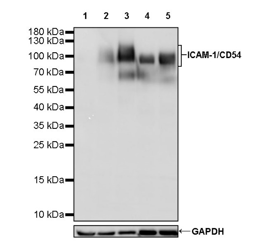
WB result of ICAM-1/CD54 Recombinant Rabbit mAb
Primary antibody: ICAM-1/CD54 Recombinant Rabbit mAb at 1/5000 dilution
Lane 1: MCF7 whole cell lysate 20 µg
Lane 2: SW480 whole cell lysate 20 µg
Lane 3: HepG2 whole cell lysate 20 µg
Lane 4: Ramos whole cell lysate 20 µg
Lane 5: Raji whole cell lysate 20 µg
Negative control: MCF7 whole cell lysate
Secondary antibody: Goat Anti-rabbit IgG, (H+L), HRP conjugated at 1/10000 dilution
Predicted MW: 58 kDa
Observed MW: 90~130 kDa
Flow cytometric analysis of human PBMC (human peripheral blood mononuclear cell) labelling ICAM-1/CD54 antibody at 1/50 (1 μg) dilution (Right) compared with a Rabbit monoclonal IgG isotype control (Left). Goat Anti - Rabbit IgG Alexa Fluor® 488 was used as the secondary antibody. Then cells were stained with CD14 - Alexa Fluor® 647 separately. Gated on total viable cells.
IHC shows positive staining in paraffin-embedded human kidney. Anti- ICAM-1/CD54 antibody was used at 1/500 dilution, followed by a HRP Polymer for Mouse & Rabbit IgG (ready to use). Counterstained with hematoxylin. Heat mediated antigen retrieval with Tris/EDTA buffer pH9.0 was performed before commencing with IHC staining protocol.
IHC shows positive staining in paraffin-embedded human spleen. Anti- ICAM-1/CD54 antibody was used at 1/500 dilution, followed by a HRP Polymer for Mouse & Rabbit IgG (ready to use). Counterstained with hematoxylin. Heat mediated antigen retrieval with Tris/EDTA buffer pH9.0 was performed before commencing with IHC staining protocol.
IHC shows positive staining in paraffin-embedded human tonsil. Anti- ICAM-1/CD54 antibody was used at 1/500 dilution, followed by a HRP Polymer for Mouse & Rabbit IgG (ready to use). Counterstained with hematoxylin. Heat mediated antigen retrieval with Tris/EDTA buffer pH9.0 was performed before commencing with IHC staining protocol.
IHC shows positive staining in paraffin-embedded human lung. Anti- ICAM-1/CD54 antibody was used at 1/500 dilution, followed by a HRP Polymer for Mouse & Rabbit IgG (ready to use). Counterstained with hematoxylin. Heat mediated antigen retrieval with Tris/EDTA buffer pH9.0 was performed before commencing with IHC staining protocol.
IHC shows positive staining in paraffin-embedded human hepatocellular carcinoma. Anti- ICAM-1/CD54 antibody was used at 1/500 dilution, followed by a HRP Polymer for Mouse & Rabbit IgG (ready to use). Counterstained with hematoxylin. Heat mediated antigen retrieval with Tris/EDTA buffer pH9.0 was performed before commencing with IHC staining protocol.
IHC shows positive staining in paraffin-embedded human lung squamous cell carcinoma. Anti- ICAM-1/CD54 antibody was used at 1/500 dilution, followed by a HRP Polymer for Mouse & Rabbit IgG (ready to use). Counterstained with hematoxylin. Heat mediated antigen retrieval with Tris/EDTA buffer pH9.0 was performed before commencing with IHC staining protocol.
 您现在的位置: