
12 months from date of receipt / reconstitution, -20 °C as supplied
| 应用 | 稀释度 |
|---|---|
| WB | 1:1000 |
| IP | 1:50 |
| ICC | 1:50 |
| ICFCM | 1:50 |
The Inhibitor of Nuclear Factor Kappa-B Kinase (IKK) is a pivotal serine/threonine kinase complex that plays a crucial role in regulating the Nuclear Factor Kappa-B (NF-κB) signaling pathway. This pathway is fundamental for controlling diverse physiological and pathological processes, including immune responses, inflammation, cell proliferation, and apoptosis. The IKK complex comprises three main subunits: IKKα (also known as IKK1), IKKβ (IKK2), and IKKγ (or NEMO/IKBKG). Each subunit contributes uniquely to the complex's structure and function. IKKα possesses kinase activity, albeit lower than IKKβ. Its precise role in NF-κB activation remains somewhat elusive, but it may be involved in specific types of NF-κB activation. IKKβ serves as the primary kinase subunit within the complex, exhibiting robust kinase activity. IKKβ is essential for activating the NF-κB pathway by phosphorylating the inhibitory protein IκB, leading to its degradation and subsequent release of NF-κB for nuclear translocation and transcriptional activation. IKK's primary function lies in the activation of the NF-κB signaling pathway. By phosphorylating specific serine residues on the IκB proteins, IKK triggers their ubiquitination and subsequent degradation by the proteasome. This process frees NF-κB from inhibition, allowing it to translocate to the nucleus and activate the transcription of target genes.
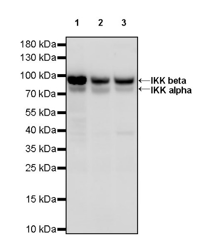
WB result of IKK alpha/beta Recombinant Rabbit mAb
Primary antibody: IKK alpha/beta Recombinant Rabbit mAb at 1/1000 dilution
Lane 1: HeLa whole cell lysate 20 µg
Lane 2: HepG2 whole cell lysate 20 µg
Lane 3: HCT 116 whole cell lysate 20 µg
Secondary antibody: Goat Anti-rabbit IgG, (H+L), HRP conjugated at 1/10000 dilution
Predicted MW: 87 kDa
Observed MW: 85, 90 kDa
WB result of IKK alpha/beta Recombinant Rabbit mAb
Primary antibody: IKK alpha/beta Recombinant Rabbit mAb at 1/1000 dilution
Lane 1: NIH/3T3 whole cell lysate 20 µg
Secondary antibody: Goat Anti-rabbit IgG, (H+L), HRP conjugated at 1/10000 dilution
Predicted MW: 87 kDa
Observed MW: 85, 90 kDa
WB result of IKK alpha/beta Recombinant Rabbit mAb
Primary antibody: IKK alpha/beta Recombinant Rabbit mAb at 1/1000 dilution
Lane 1: C6 whole cell lysate 20 µg
Secondary antibody: Goat Anti-rabbit IgG, (H+L), HRP conjugated at 1/10000 dilution
Predicted MW: 87 kDa
Observed MW: 85, 90 kDa
Flow cytometric analysis of 4% PFA fixed 90% methanol permeabilized HeLa (Human cervix adenocarcinoma epithelial cell) labelling IKK alpha/beta antibody at 1/50 dilution (1 μg)/ (Red) compared with a Rabbit monoclonal IgG (Black) isotype control and an unlabelled control (cells without incubation with primary antibody and secondary antibody) (Blue). Goat Anti - Rabbit IgG Alexa Fluor® 488 was used as the secondary antibody.
IKK alpha/beta Rabbit mAb at 1/50 dilution (1 µg) immunoprecipitating IKK alpha/beta in 0.4 mg HeLa whole cell lysate.
Western blot was performed on the immunoprecipitate using IKK alpha/beta Rabbit mAb at 1/1000 dilution.
Secondary antibody (HRP) for IP was used at 1/1000 dilution.
Lane 1: HeLa whole cell lysate 20 µg (Input)
Lane 2: IKK alpha/beta Rabbit mAb IP in HeLa whole cell lysate
Lane 3: Rabbit monoclonal IgG IP in HeLa whole cell lysate
Predicted MW: 87 kDa
Observed MW: 85, 90 kDa
ICC shows positive staining in HeLa cells. Anti- IKK alpha beta antibody was used at 1/50 dilution (Green) and incubated overnight at 4°C. Goat polyclonal Antibody to Rabbit IgG - H&L (Alexa Fluor® 488) was used as secondary antibody at 1/1000 dilution. The cells were fixed with 100% ice-cold methanol and permeabilized with 0.1% PBS-Triton X-100. Nuclei were counterstained with DAPI (Blue). Counterstain with tubulin (Red).
 您现在的位置: