
PBS, 40% Glycerol, 0.05% BSA, 0.03% Proclin 300
12 months from date of receipt / reconstitution, -20 °C as supplied
| 应用 | 稀释度 |
|---|---|
| WB | 1:1000 |
| IP | 1:50 |
| ICC | 1:500 |
| ICFCM | 1:50 |
Jun N-terminal Kinase 2, also known as JNK2, is a member of the mitogen-activated protein kinase (MAPK) family, specifically belonging to the subgroup of stress-activated protein kinases (SAPKs). JNK2 plays a pivotal role in the regulation of cell survival, apoptosis, proliferation, differentiation, and inflammatory responses. Upon activation by upstream signaling molecules, JNK2 phosphorylates various transcription factors, particularly members of the c-Jun family (including c-Jun itself), as well as ATF2, p53, and others. This phosphorylation event modulates the transcriptional activity of these factors, ultimately influencing the expression of genes involved in the aforementioned cellular processes. The activation of JNK2 is a complex process that involves the phosphorylation of specific threonine and tyrosine residues within its activation loop by MAPK kinases (MKKs), primarily MKK4 and MKK7. This phosphorylation event triggers a conformational change in JNK2, allowing it to become fully active and capable of phosphorylating its downstream substrates. The upstream signaling pathways that lead to JNK2 activation are diverse and include pathways activated by TNF-α, IL-1, and other inflammatory cytokines, as well as pathways activated by environmental stresses such as UV radiation and oxidative stress.
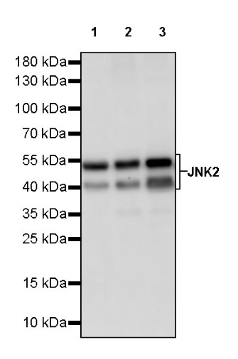
WB result of JNK2 Recombinant Rabbit mAb
Primary antibody: JNK2 Recombinant Rabbit mAb at 1/1000 dilution
Lane 1: HeLa whole cell lysate 20 µg
Lane 2: K562 whole cell lysate 20 µg
Lane 3: MCF7 whole cell lysate 20 µg
Secondary antibody: Goat Anti-rabbit IgG, (H+L), HRP conjugated at 1/10000 dilution
Predicted MW: 48 kDa
Observed MW: 46, 54 kDa
WB result of JNK2 Recombinant Rabbit mAb
Primary antibody: JNK2 Recombinant Rabbit mAb at 1/1000 dilution
Lane 1: NIH/3T3 whole cell lysate 20 µg
Secondary antibody: Goat Anti-rabbit IgG, (H+L), HRP conjugated at 1/10000 dilution
Predicted MW: 48 kDa
Observed MW: 46, 54 kDa
WB result of JNK2 Recombinant Rabbit mAb
Primary antibody: JNK2 Recombinant Rabbit mAb at 1/1000 dilution
Lane 1: PC-12 whole cell lysate 20 µg
Secondary antibody: Goat Anti-rabbit IgG, (H+L), HRP conjugated at 1/10000 dilution
Predicted MW: 48 kDa
Observed MW: 46, 54 kDa
Flow cytometric analysis of 4% PFA fixed 90% methanol permeabilized HeLa (Human cervix adenocarcinoma epithelial cell) labelling JNK2 antibody at 1/50 dilution (1 μg)/ (Red) compared with a Rabbit monoclonal IgG (Black) isotype control and an unlabelled control (cells without incubation with primary antibody and secondary antibody) (Blue). Goat Anti - Rabbit IgG Alexa Fluor® 488 was used as the secondary antibody.
Flow cytometric analysis of 4% PFA fixed 90% methanol permeabilized NIH/3T3 (Mouse embryonic fibroblast) labelling JNK2 antibody at 1/50 dilution (1 μg)/ (Red) compared with a Rabbit monoclonal IgG (Black) isotype control and an unlabelled control (cells without incubation with primary antibody and secondary antibody) (Blue). Goat Anti - Rabbit IgG Alexa Fluor® 488 was used as the secondary antibody.
JNK2 Rabbit mAb at 1/50 dilution (1 µg) immunoprecipitating JNK2 in 0.4 mg HeLa whole cell lysate.
Western blot was performed on the immunoprecipitate using JNK2 Rabbit mAb at 1/1000 dilution.
Secondary antibody (HRP) for IP was used at 1/1000 dilution.
Lane 1: HeLa whole cell lysate 20 µg (Input)
Lane 2: JNK2 Rabbit mAb IP in HeLa whole cell lysate
Lane 3: Rabbit monoclonal IgG IP in HeLa whole cell lysate
Predicted MW: 48 kDa
Observed MW: 46, 54 kDa
ICC shows positive staining in HeLa cells. Anti-JNK2 antibody was used at 1/500 dilution (Green) and incubated overnight at 4°C. Goat polyclonal Antibody to Rabbit IgG - H&L (Alexa Fluor® 488) was used as secondary antibody at 1/1000 dilution. The cells were fixed with 100% ice-cold methanol and permeabilized with 0.1% PBS-Triton X-100. Nuclei were counterstained with DAPI (Blue). Counterstain with tubulin (Red).
ICC shows positive staining in NIH/3T3 cells. Anti-JNK2 antibody was used at 1/500 dilution (Green) and incubated overnight at 4°C. Goat polyclonal Antibody to Rabbit IgG - H&L (Alexa Fluor® 488) was used as secondary antibody at 1/1000 dilution. The cells were fixed with 100% ice-cold methanol and permeabilized with 0.1% PBS-Triton X-100. Nuclei were counterstained with DAPI (Blue). Counterstain with tubulin (Red).
 您现在的位置: