
12 months from date of receipt / reconstitution, -20 °C as supplied
| 应用 | 稀释度 |
|---|---|
| WB | 1:1000 |
| ICC | 1:500 |
| ICFCM | 1:500 |
As an E3 ubiquitin-protein ligase, MDM2 is involved in the ubiquitination and subsequent degradation of various proteins, primarily targeting the tumor suppressor protein p53/TP53. By mediating p53's degradation, MDM2 regulates cell cycle progression, apoptosis, and DNA damage repair. In normal cells, MDM2 maintains a delicate balance with p53, jointly modulating cell cycle arrest, apoptosis, and DNA repair to ensure cellular homeostasis. In cancer, MDM2 is frequently overexpressed, leading to increased degradation of p53, thereby inhibiting apoptosis and cell cycle arrest, contributing to tumorigenesis. MDM2 overexpression is also associated with tumor microenvironment remodeling and immune evasion. Furthermore, MDM2 exerts bidirectional effects on CD8+ T cells, CD4+ T cells, and other immune cells like NK cells, DCs, Tregs, and TAMs, shaping the tumor immune microenvironment.
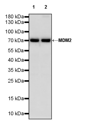
WB result of MDM2 Recombinant Rabbit mAb
Primary antibody: MDM2 Recombinant Rabbit mAb at 1/1000 dilution
Lane 1: HeLa whole cell lysate 20 µg
Lane 2: SK-OV-3 whole cell lysate 20 µg
Secondary antibody: Goat Anti-rabbit IgG, (H+L), HRP conjugated at 1/10000 dilution
Predicted MW: 55 kDa
Observed MW: 75 kDa
WB result of MDM2 Recombinant Rabbit mAb
Primary antibody: MDM2 Recombinant Rabbit mAb at 1/1000 dilution
Lane 1: RAW264.7 whole cell lysate 20 µg
Secondary antibody: Goat Anti-rabbit IgG, (H+L), HRP conjugated at 1/10000 dilution
Predicted MW: 55 kDa
Observed MW: 75 kDa
Flow cytometric analysis of 4% PFA fixed 90% methanol permeabilized HeLa (Human cervix adenocarcinoma epithelial cell) labelling MDM2 antibody at 1/500 dilution (0.1 μg)/ (Red) compared with a Rabbit monoclonal IgG (Black) isotype control and an unlabelled control (cells without incubation with primary antibody and secondary antibody) (Blue). Goat Anti - Rabbit IgG Alexa Fluor® 488 was used as the secondary antibody.
ICC analysis of U-2 OS cells treated with Nutlin 3a (10 μM, 24 hr) (top panel) and U-2 OS cells untreated with Nutlin 3a (10 μM, 24 hr) (below panel). Anti-MDM2 antibody was used at 1/500 dilution (Green) and incubated overnight at 4°C. Goat polyclonal Antibody to Rabbit IgG - H&L (Alexa Fluor® 488) was used as secondary antibody at 1/1000 dilution. The cells were fixed with 4% PFA and permeabilized with 0.1% PBS-Triton X-100. Nuclei were counterstained with DAPI (Blue). Counterstain with tubulin (Red).
 您现在的位置: