
12 months from date of receipt / reconstitution, -20 °C as supplied
| 应用 | 稀释度 |
|---|---|
| WB | 1:1000 |
| IHC | 1:1000-1:4000 |
| ICC | 1:500 |
The transcription factor Pax5 is essential for commitment of lymphoid progenitors to the B lymphocyte lineage. Pax5 fulfils a dual role by repressing B lineage 'inappropriate' genes and simultaneously activating B lineage–specific genes. This transcriptional reprogramming restricts the broad signaling capacity of uncommitted progenitors to the B cell pathway, regulates cell adhesion and migration, induces VH-DJH recombination, facilitates (pre-)B cell receptor signaling and promotes development to the mature B cell stage. Conditional Pax5 inactivation in early and late B lymphocytes revealed an essential role for Pax5 in controlling the identity and function of B cells throughout B lymphopoiesis. PAX5 has also been implicated in human B cell malignancies, as it is deregulated by chromosomal translocations in a subset of acute lymphoblastic leukemias and non-Hodgkin lymphomas.
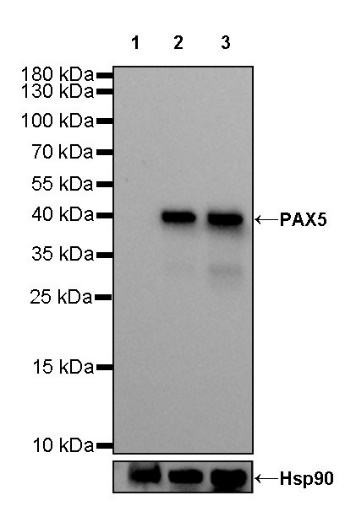
WB result of PAX5 Rat mAb Primary antibody: PAX5 Rat mAb at 1/1000 dilution Lane 1: Jurkat whole cell lysate 20 µg Lane 2: Ramos whole cell lysate 20 µg Lane 3: Daudi whole cell lysate 20 µg Negative control: Jurkat whole cell lysate Secondary antibody: Goat Anti-Rat IgG, (H+L), HRP conjugated at 1/10000 dilution Predicted MW: 42 kDa Observed MW: 42 kDa
IHC shows positive staining in paraffin-embedded human tonsil. Anti-PAX5 antibody was used at 1/4000 dilution, followed by a HRP Polymer for Goat Anti-Rat IgG (H+L). Counterstained with hematoxylin. Heat mediated antigen retrieval with Tris/EDTA buffer pH9.0 was performed before commencing with IHC staining protocol.
IHC shows positive staining in paraffin-embedded human diffuse large B-cell lymphoma. Anti-PAX5 antibody was used at 1/4000 dilution, followed by a HRP Polymer for Goat Anti-Rat IgG (H+L). Counterstained with hematoxylin. Heat mediated antigen retrieval with Tris/EDTA buffer pH9.0 was performed before commencing with IHC staining protocol.
Negative control: IHC shows negative staining in paraffin-embedded human NK/T-cell lymphoma. Anti-PAX5 antibody was used at 1/4000 dilution, followed by a HRP Polymer for Goat Anti-Rat IgG (H+L). Counterstained with hematoxylin. Heat mediated antigen retrieval with Tris/EDTA buffer pH9.0 was performed before commencing with IHC staining protocol.
IHC shows positive staining in paraffin-embedded mouse spleen. Anti-PAX5 antibody was used at 1/4000 dilution, followed by a HRP Polymer for Goat Anti-Rat IgG (H+L). Counterstained with hematoxylin. Heat mediated antigen retrieval with Tris/EDTA buffer pH9.0 was performed before commencing with IHC staining protocol.
IHC shows positive staining in paraffin-embedded rat spleen. Anti-PAX5 antibody was used at 1/4000 dilution, followed by a HRP Polymer for Goat Anti-Rat IgG (H+L). Counterstained with hematoxylin. Heat mediated antigen retrieval with Tris/EDTA buffer pH9.0 was performed before commencing with IHC staining protocol.
ICC shows positive staining in Ramos cells. Anti-PAX5 antibody was used at 1/500 dilution (Green) and incubated overnight at 4°C. Goat polyclonal Antibody to Rat IgG - H&L (Alexa Fluor® 488) was used as secondary antibody at 1/1000 dilution. The cells were fixed with 4% PFA and permeabilized with 0.1% PBS-Triton X-100. Nuclei were counterstained with DAPI.
Negative control: ICC shows negative staining in Jurkat cells. Anti-PAX5 antibody was used at 1/500 dilution and incubated overnight at 4°C. Goat polyclonal Antibody to Rat IgG - H&L (Alexa Fluor® 488) was used as secondary antibody at 1/1000 dilution. The cells were fixed with 4% PFA and permeabilized with 0.1% PBS-Triton X-100. Nuclei were counterstained with DAPI.
 您现在的位置: