
| 应用 | 稀释度 |
|---|---|
| IHC-P | 1:2000 |
| ICC | 1:250 |
| WB | 1:1000 |
| IF | 1:1000 |
Calponin-1 is a 34 kDa actin-binding protein with regions of sequence homology to cardiac troponin I and T. It is located in the cytoskeleton and contractile apparatus of differentiated smooth muscle cells. There are 3 isoforms of calponin: calponin-1 (h1 or basic), calponin-2 (h2 or neutral), and calponin-3 (h3 or acidic). Calponin-1 is implicated in the regulation of smooth muscle contraction by mediating intracellular signaling responses to some vasoconstrictors by acting as a contractile scaffold protein. This mechanism is based on results in aortic smooth muscle cells stimulated with phenylephrine, which showed calponin-1 connecting protein kinase C and ERK1/2 pathways to promote contractility.

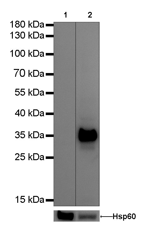
WB result of Calponin-1 Rabbit mAb
Primary antibody : Calponin-1 Rabbit mAb at 1/1000 dilution
Lane 1: 293-T whole cell lysate 20 µg
Lane 2: mouse bladder lysate 20 µg
Negative control: 293-T whole cell lysate
Secondary antibody: Goat Anti-Rabbit IgG, (H+L), HRP conjugated at 1/10000 dilution
Predicted MW: 34 kDa
Observed MW: 34 kDa
Exposure time: 0.3s
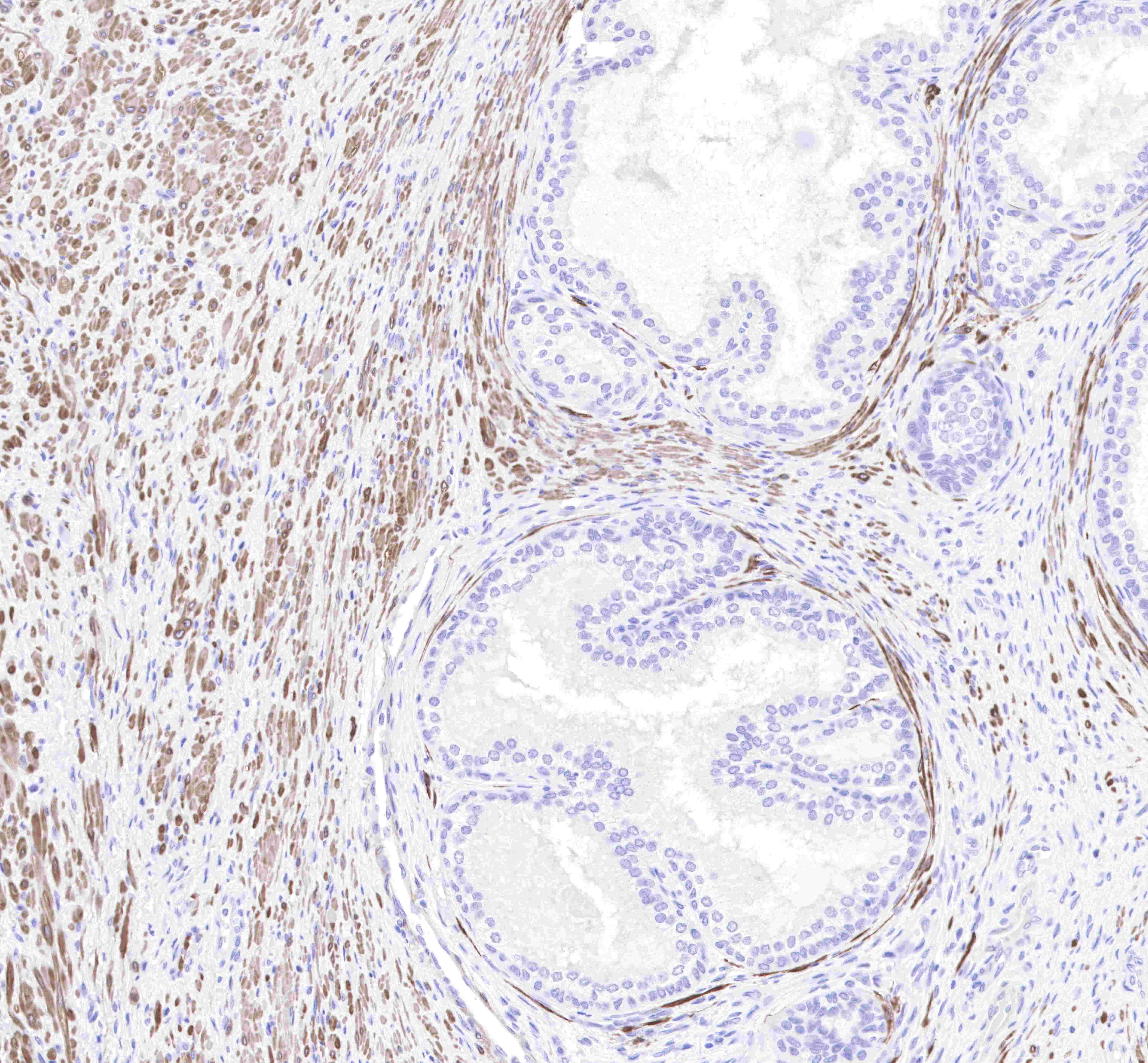
IHC shows positive staining in paraffin-embedded human prostate. Anti-Calponin-1 antibody was used at 1/2000 dilution, followed by a Goat Anti-Rabbit IgG H&L (HRP) ready to use. Counterstained with hematoxylin. Heat mediated antigen retrieval with Tris/EDTA buffer pH9.0 was performed before commencing with IHC staining protocol.
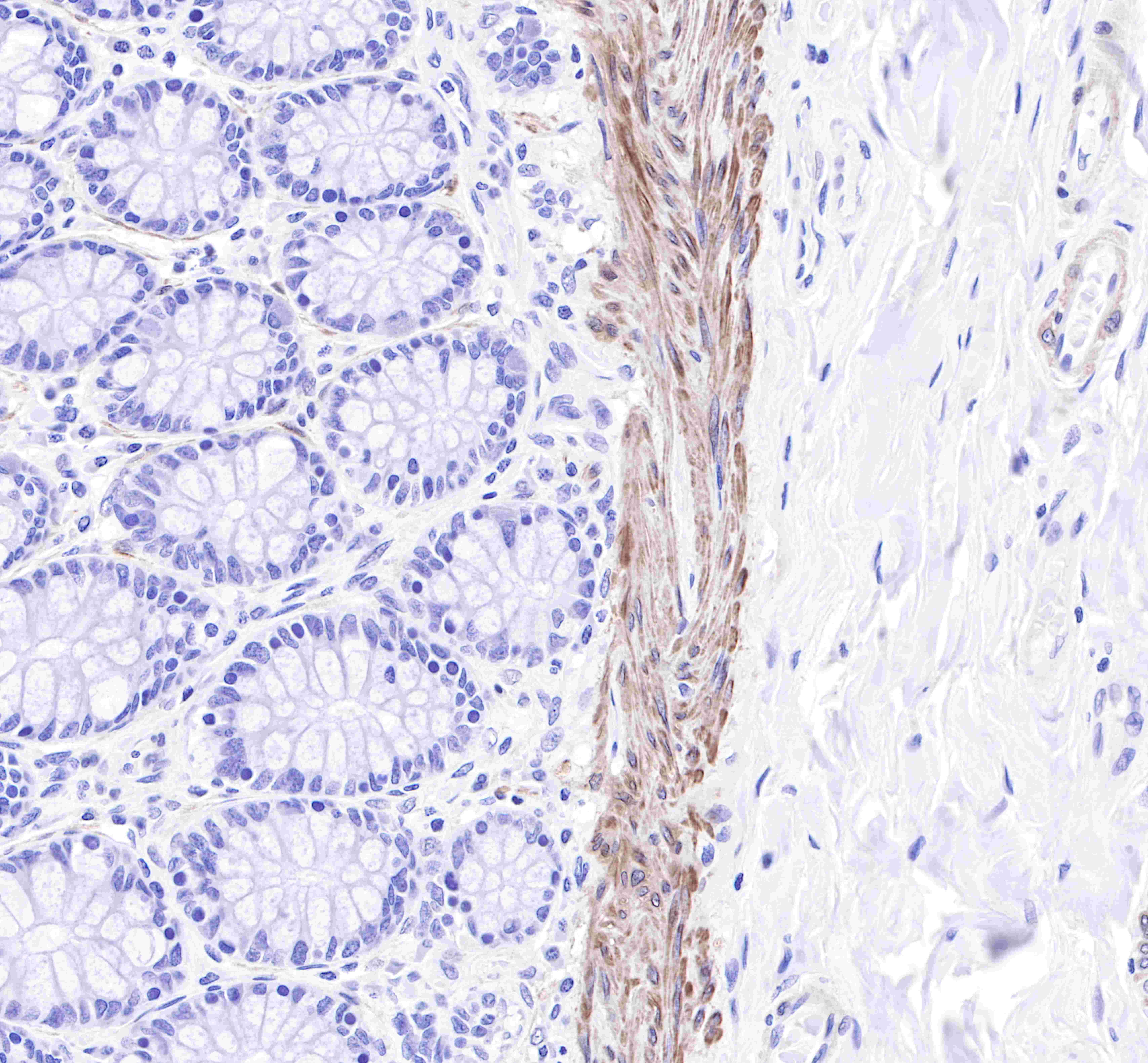
IHC shows positive staining in paraffin-embedded human colon. Anti-Calponin-1 antibody was used at 1/2000 dilution, followed by a Goat Anti-Rabbit IgG H&L (HRP) ready to use. Counterstained with hematoxylin. Heat mediated antigen retrieval with Tris/EDTA buffer pH9.0 was performed before commencing with IHC staining protocol.
IHC shows positive staining in paraffin-embedded human cervix cancer. Anti-Calponin-1 antibody was used at 1/2000 dilution, followed by a Goat Anti-Rabbit IgG H&L (HRP) ready to use. Counterstained with hematoxylin. Heat mediated antigen retrieval with Tris/EDTA buffer pH9.0 was performed before commencing with IHC staining protocol.
IHC shows positive staining in paraffin-embedded human breast. Anti-Calponin-1 antibody was used at 1/2000 dilution, followed by a Goat Anti-Rabbit IgG H&L (HRP) ready to use. Counterstained with hematoxylin. Heat mediated antigen retrieval with Tris/EDTA buffer pH9.0 was performed before commencing with IHC staining protocol.
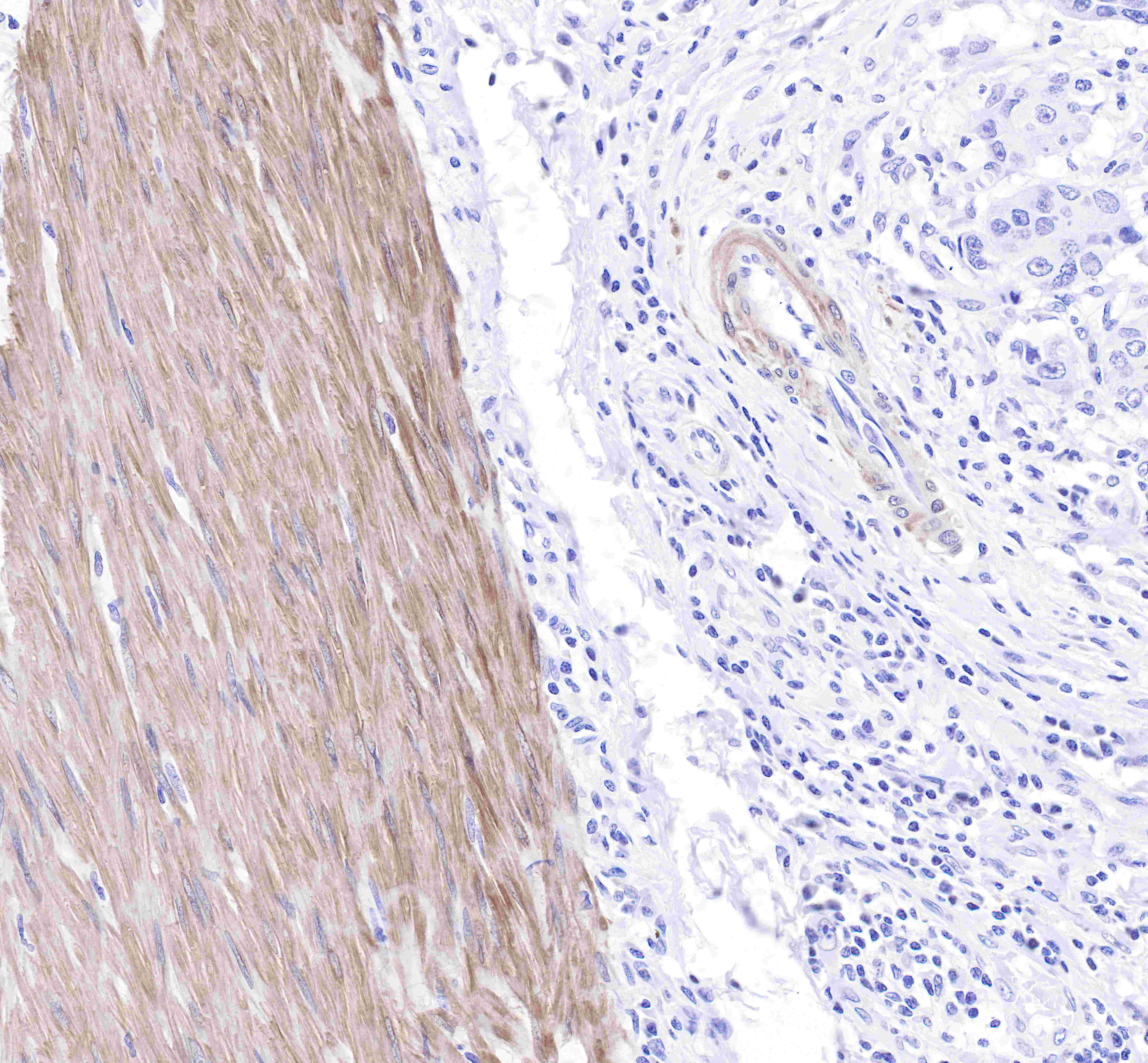
IHC shows positive staining in paraffin-embedded mouse stomach. Anti-Calponin-1 antibody was used at 1/2000 dilution, followed by a Goat Anti-Rabbit IgG H&L (HRP) ready to use. Counterstained with hematoxylin. Heat mediated antigen retrieval with Tris/EDTA buffer pH9.0 was performed before commencing with IHC staining protocol.
IHC shows positive staining in paraffin-embedded rat colon. Anti-Calponin-1 antibody was used at 1/2000 dilution, followed by a Goat Anti-Rabbit IgG H&L (HRP) ready to use. Counterstained with hematoxylin. Heat mediated antigen retrieval with Tris/EDTA buffer pH9.0 was performed before commencing with IHC staining protocol.
ICC shows cytoplasm staining in Panc-1 cell.
Anti-Calponin-1 antibody was used at 1/250 dilution and incubated overnight at 4°C.
Goat polyclonal Antibody to Rabbit IgG - H&L (Alexa Fluor® 488) was used as secondary antibody at 1/1000 dilution.
Cells were fixed with 4% paraformaldehyde and permeabilized with 0.1% Triton X-100.
Nuclei were counterstained with DAPI.
ICC shows negative staining in 293T cells.
Anti-Calponin-1 antibody was used at 1/250 dilution and incubated overnight at 4°C.
Goat polyclonal Antibody to Rabbit IgG - H&L (Alexa Fluor® 488) was used as secondary antibody at 1/1000 dilution.
Cells were fixed with 100% Methanol and permeabilized with 0.1% Triton X-100.
Nuclei were counterstained with DAPI.
IF shows positive staining in paraffin-embedded human breast. Anti-Calponin-1 antibody was used at 1/1000 dilution (magenta) and incubated overnight at 4°C. Goat anti-Rabbit IgG (H+L) (Alexa Fluor® 647 Conjugate) (S0B4005) was used as secondary antibody at 1/1000 dilution. Counterstained with DAPI (Blue). Heat mediated antigen retrieval with EDTA buffer pH9.0 was performed before commencing with IF staining protocol.
IF shows positive staining in paraffin-embedded human prostate. Anti-Calponin-1 antibody was used at 1/1000 dilution (magenta) and incubated overnight at 4°C. Goat anti-Rabbit IgG (H+L) (Alexa Fluor® 647 Conjugate) (S0B4005)was used as secondary antibody at 1/1000 dilution. Counterstained with DAPI (Blue). Heat mediated antigen retrieval with EDTA buffer pH9.0 was performed before commencing with IF staining protocol.
IF shows positive staining in paraffin-embedded mouse stomach . Anti-Calponin-1 antibody was used at 1/1000 dilution (magenta) and incubated overnight at 4°C. Goat anti-Rabbit IgG (H+L) (Alexa Fluor® 647 Conjugate) (S0B4005)was used as secondary antibody at 1/1000 dilution. Counterstained with DAPI (Blue). Heat mediated antigen retrieval with EDTA buffer pH9.0 was performed before commencing with IF staining protocol.

IF shows positive staining in paraffin-embedded rat stomach. Anti-Calponin-1 antibody was used at 1/1000 dilution (magenta) and incubated overnight at 4°C.Goat anti-Rabbit IgG (H+L) (Alexa Fluor® 647 Conjugate) (S0B4005) was used as secondary antibody at 1/1000 dilution. Counterstained with DAPI (Blue). Heat mediated antigen retrieval with EDTA buffer pH9.0 was performed before commencing with IF staining protocol.
 您现在的位置: