

12 months from date of receipt / reconstitution, -20 °C as supplied
| 应用 | 稀释度 |
|---|---|
| ICC | 1:500 |
| WB | 1:1000 |
| IP | 1:25 |
| IHC-P | 1:500-1:1000 |
| IF | 1:500 |
CD34 is a transmembrane phosphoglycoprotein protein encoded by the CD34 gene. CD34 derives its name from the cluster of differentiation protocol that identifies cell surface antigens. CD34 was first described on hematopoietic stem cells independently by Civin et al. and Tindle et al, as a cell surface glycoprotein and functions as a cell-cell adhesion factor. It may also mediate the attachment of hematopoietic stem cells to bone marrow extracellular matrix or directly to stromal cells. Clinically, it is associated with the selection and enrichment of hematopoietic stem cells for bone marrow transplants. Due to these historical and clinical associations, CD34 expression is almost ubiquitously related to hematopoietic cells.
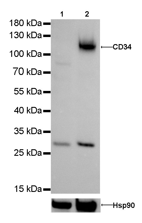
WB result of CD34 Rabbit mAb
Primary antibody : CD34 Rabbit mAb at 1/1000 dilution
Lane 1: Hela whole cell lysate 20 µg
Lane 2: TF-1 whole cell lysate 20 µg
Negative control: Hela whole cell lysate
Secondary antibody: Goat Anti-Rabbit IgG, (H+L), HRP conjugated at 1/10000 dilution
Predicted MW: 120 kDa
Observed MW: 120 kDa
Exposure time: 30s
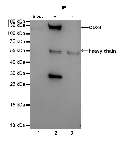
CD34 Rabbit mAb at 1/25 dilution (2µg) immunoprecipitating CD34 in 0.4mg TF-1 whole cell lysate. Western blot was performed on the immunoprecipitate using CD34 Rabbit mAb at 1/1000 dilution. Secondary antibody (HRP) for IP was used at 1/400 dilution.
Lane 1: TF-1 whole cell lysate 10µg (input)
Lane 2: CD34 Rabbit mAb IP in TF-1 whole cell lysate
Lane 3: Rabbit monoclonal IgG IP in TF-1 whole cell lysate
Predicted MW: 120 kDa
Observed MW: 120 kDa
Exposure time: 180s
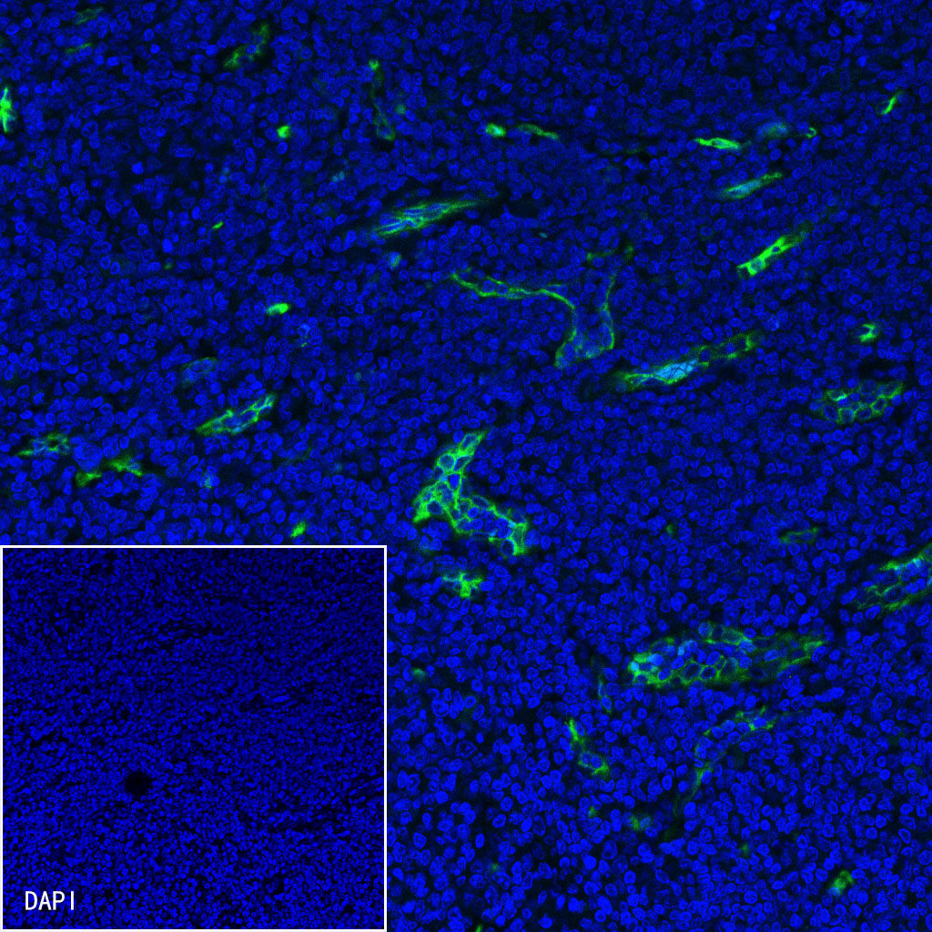
IHC shows positive staining in paraffin-embedded human tonsil.
Anti-CD34 antibody was used at 1/2000 dilution, followed by a Goat Anti-Rabbit IgG H&L (HRP) ready to use.
Counterstained with hematoxylin.
Heat mediated antigen retrieval with Tris/EDTA buffer pH9.0 was performed before commencing with IHC staining protocol.
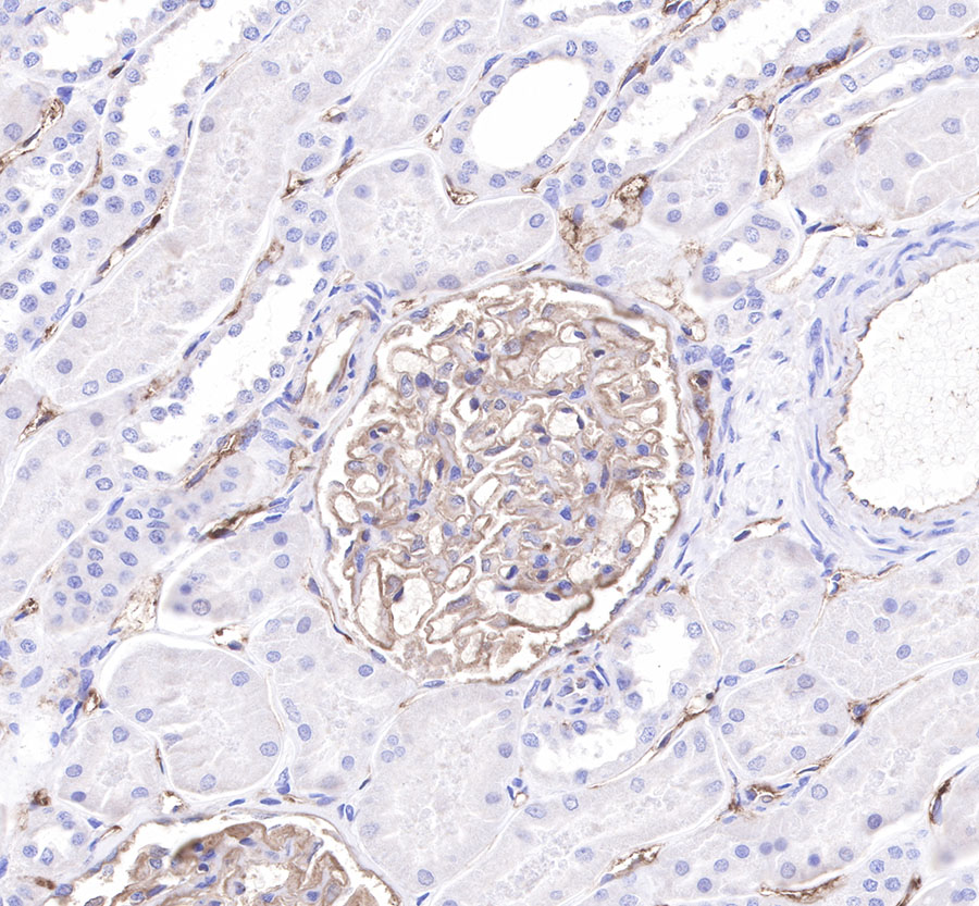
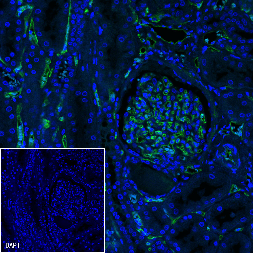
IHC shows positive staining in paraffin-embedded paraffin-embedded human kidney.
Anti-CD34 antibody was used at 1/2000 dilution, followed by a Goat Anti-Rabbit IgG H&L (HRP) ready to use.
Counterstained with hematoxylin.
Heat mediated antigen retrieval with Tris/EDTA buffer pH9.0 was performed before commencing with IHC staining protocol.
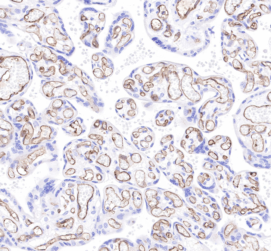
IHC shows positive staining in paraffin-embedded human placenta.
Anti-CD34 antibody was used at 1/2000 dilution, followed by a Goat Anti-Rabbit IgG H&L (HRP) ready to use.
Counterstained with hematoxylin.
Heat mediated antigen retrieval with Tris/EDTA buffer pH9.0 was performed before commencing with IHC staining protocol.
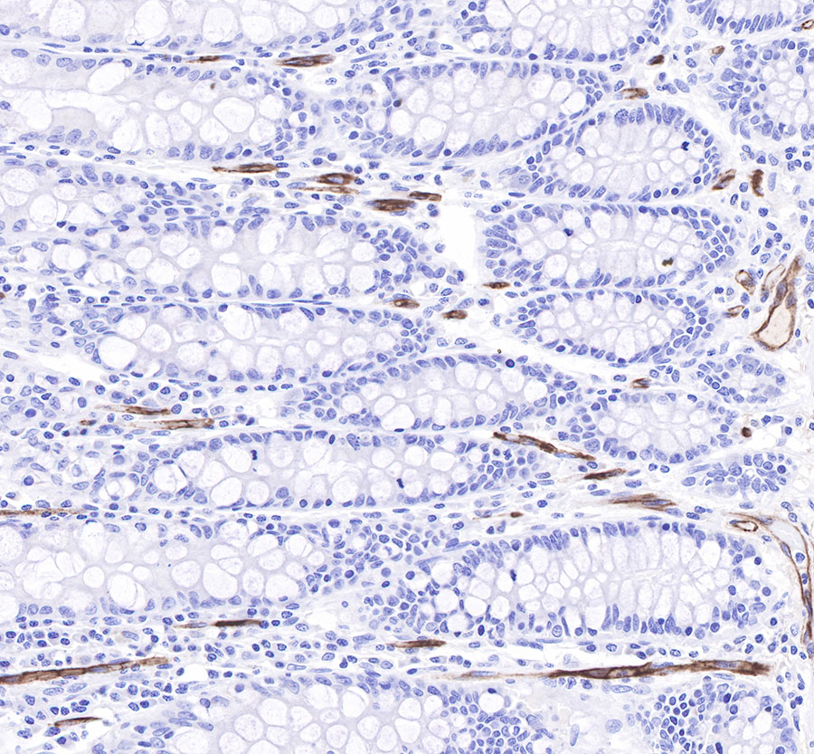
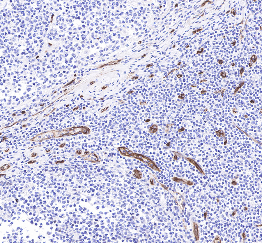
IHC shows positive staining in paraffin-embedded human colon.
Anti-CD34 antibody was used at 1/2000 dilution, followed by a Goat Anti-Rabbit IgG H&L (HRP) ready to use.
Counterstained with hematoxylin.
Heat mediated antigen retrieval with Tris/EDTA buffer pH9.0 was performed before commencing with IHC staining protocol.
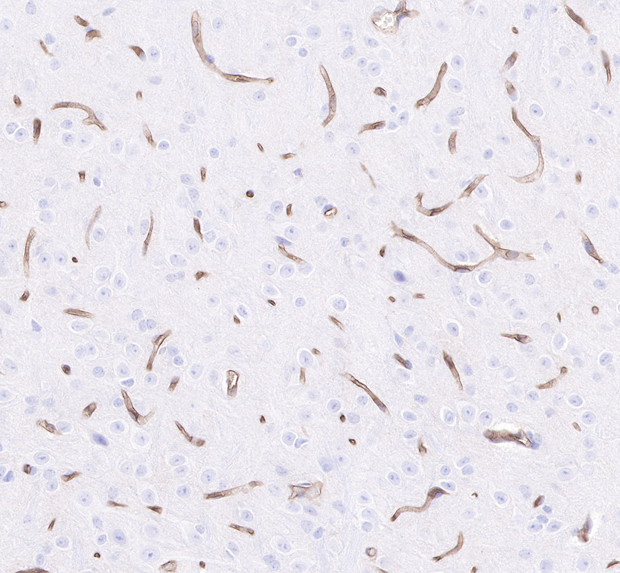
IHC shows positive staining in paraffin-embedded mouse brain.
Anti-CD34 antibody was used at 1/2000 dilution, followed by a Goat Anti-Rabbit IgG H&L (HRP) ready to use.
Counterstained with hematoxylin.
Heat mediated antigen retrieval with Tris/EDTA buffer pH9.0 was performed before commencing with IHC staining protocol.
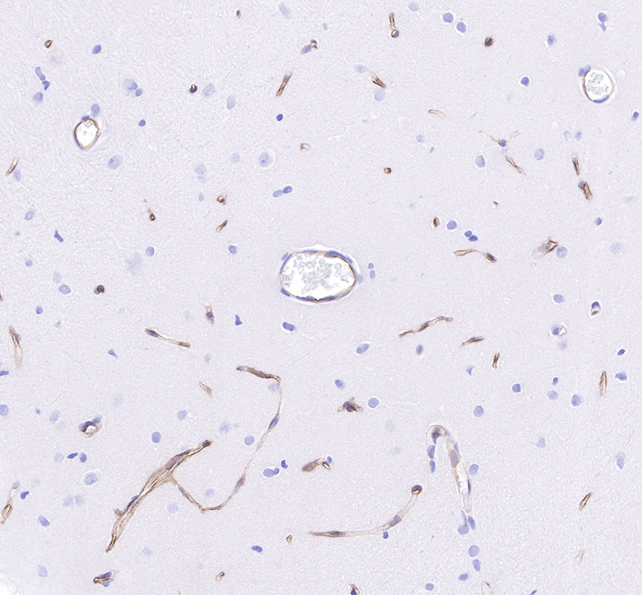
IHC shows positive staining in paraffin-embedded rat brain.
Anti-CD34 antibody was used at 1/2000 dilution, followed by a Goat Anti-Rabbit IgG H&L (HRP) ready to use.
Counterstained with hematoxylin.
Heat mediated antigen retrieval with Tris/EDTA buffer pH9.0 was performed before commencing with IHC staining protocol.
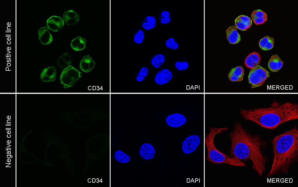
ICC shows positive staining in TF-1 cells (top panel) and negative staining in HeLa cells (below panel). Anti-CD34 antibody was used at 1/500 dilution (Green) and incubated overnight at 4°C. Goat polyclonal Antibody to Rabbit IgG - H&L (Alexa Fluor® 488) was used as secondary antibody at 1/1000 dilution. The cells were fixed with 4% PFA and permeabilized with 0.1% PBS-Triton X-100. Nuclei were counterstained with DAPI (Blue). Counterstain with tubulin (Red).
ICC shows positive staining in TF-1 cells (top panel) and negative staining in HeLa cells (below panel). Anti-CD34 antibody was used at 1/500 dilution (Green) and incubated overnight at 4°C. Goat polyclonal Antibody to Rabbit IgG - H&L (Alexa Fluor® 488) was used as secondary antibody at 1/1000 dilution. The cells were fixed with 4% PFA and permeabilized with 0.1% PBS-Triton X-100. Nuclei were counterstained with DAPI (Blue). Counterstain with tubulin (Red).
IF shows positive staining in paraffin-embedded human tonsil. Anti-CD34 antibody was used at 1/500 dilution (Green) and incubated overnight at 4°C. Goat polyclonal Antibody to Rabbit IgG - H&L (Alexa Fluor® 488) was used as secondary antibody at 1/1000 dilution. Counterstained with DAPI (Blue). Heat mediated antigen retrieval with EDTA buffer pH9.0 was performed before commencing with IF staining protocol.
IF shows positive staining in paraffin-embedded human kidney. Anti-CD34 antibody was used at 1/500 dilution (Green) and incubated overnight at 4°C. Goat polyclonal Antibody to Rabbit IgG - H&L (Alexa Fluor® 488) was used as secondary antibody at 1/1000 dilution. Counterstained with DAPI (Blue). Heat mediated antigen retrieval with EDTA buffer pH9.0 was performed before commencing with IF staining protocol.
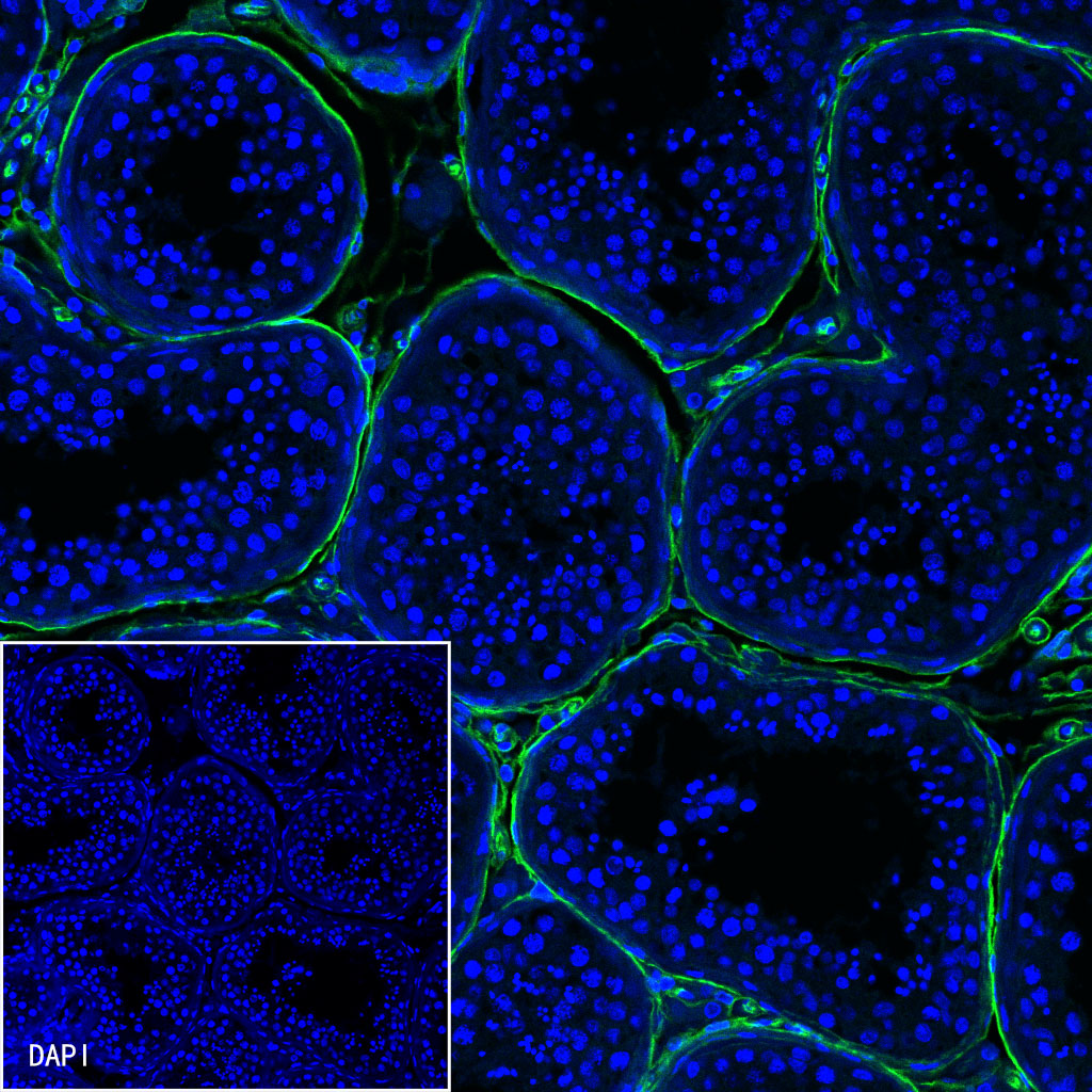
IF shows positive staining in paraffin-embedded human testis. Anti-CD34 antibody was used at 1/500 dilution (Green) and incubated overnight at 4°C. Goat polyclonal Antibody to Rabbit IgG - H&L (Alexa Fluor® 488) was used as secondary antibody at 1/1000 dilution. Counterstained with DAPI (Blue). Heat mediated antigen retrieval with EDTA buffer pH9.0 was performed before commencing with IF staining protocol.
 您现在的位置: