
93kDa
12 months from date of receipt / reconstitution, -20 °C as supplied
| 应用 | 稀释度 |
|---|---|
| ICFCM | 1:50 |
| IHC-P | 1:2000 |
| ICC | 1:500 |
| WB | 1:1000 |
| IF | 1:200-1:1000 |
VIL1 is a gastrointestinal-related cytoskeletal protein that is associated with the microfilament bundles of brush border microvilli. A major structural component of the brush border cytoskeleton, VIL1 binds actin in a calcium-dependent manner. Under normal physiological conditions, villin1 is expressed in epithelial cells of the intestinal mucosa, gall bladder, renal proximal tubules and ductuli efferentes of the testis. Wang et al. report VIL1 to be an epithelial cell-specific anti-apoptotic protein, and to have an important function in regulating actin dynamics, cell morphology, epithelial-to-mesenchymal transitions, cell migration and cell survival. Furthermore, the overexpression of villin has been reported in many diseases, such as gastrointestinal neuroendocrine tumors and colon cancer; and in lung cancers, villin1 is reported to be a useful marker to distinguish large cell neuroendocrine carcinomas from squamous cell carcinomas from the sera of patients with lung cancer (PMID:22530999).

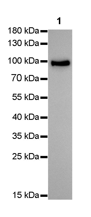
WB result of Villin Rabbit mAb
Primary antibody : Villin Rabbit mAb at 1/1000 dilution
Lane 1 : HepG2 whole cell lysate 10 µg
Secondary antibody: Goat Anti-Rabbit IgG, (H+L), HRP conjugated at 1/10000 dilution
Predicted MW: 93 kDa
Observed MW: 93 kDa
Exposure time: 15 seconds
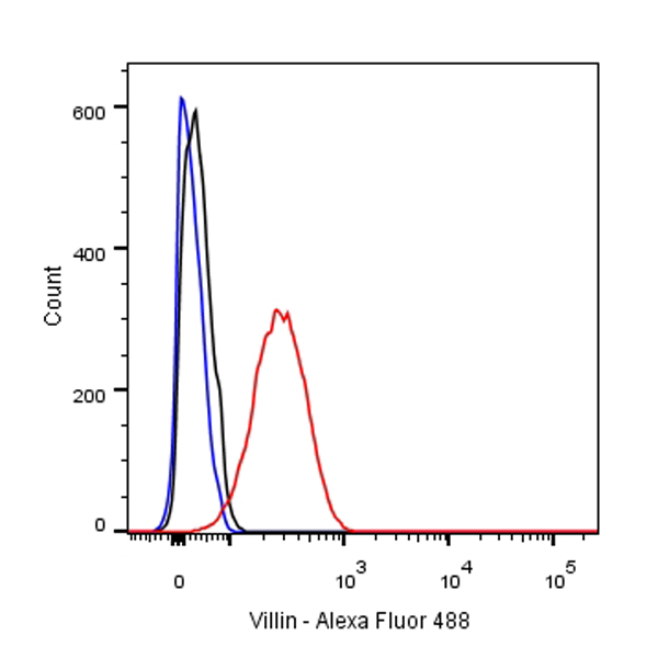
Flow cytometric analysis of HepG2 cells labelling Villin antibody at 1/50 (1ug) dilution/ (red) compared with a Rabbit monoclonal IgG (Black) isotype control and an unlabelled control (cells without incubation with primary antibody and secondary antibody) (Blue). Goat Anti-Rabbit IgG Alexa Fluor® 488 was used as the secondary antibody.
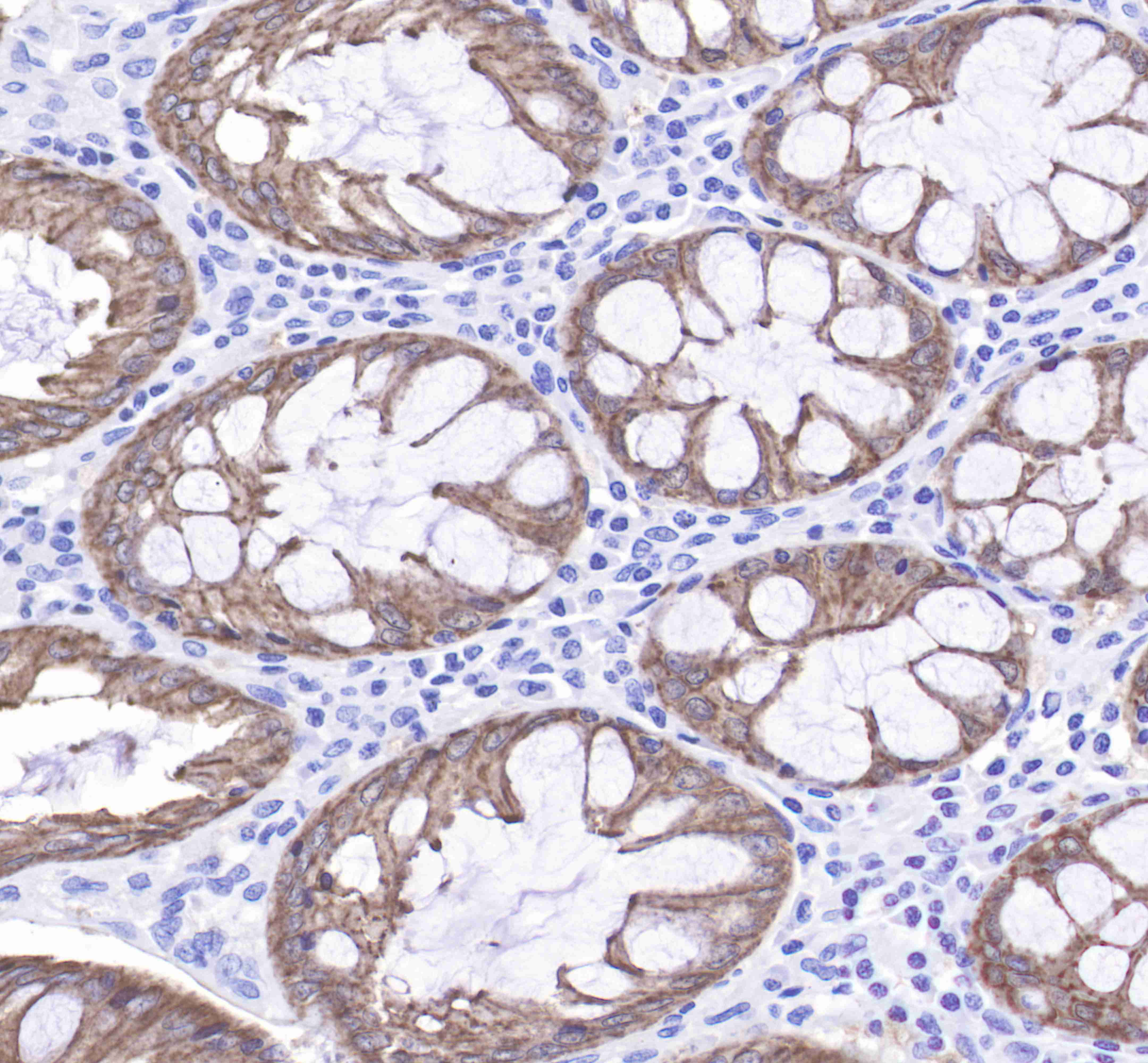
IHC shows positive staining in paraffin-embedded human colon.
Anti-Villin antibody was used at 1/2000 dilution, followed by a Goat Anti-Rabbit IgG H&L (HRP) ready to use.
Counterstained with hematoxylin.
Heat mediated antigen retrieval with Tris/EDTA buffer pH9.0 was performed before commencing with IHC staining protocol.
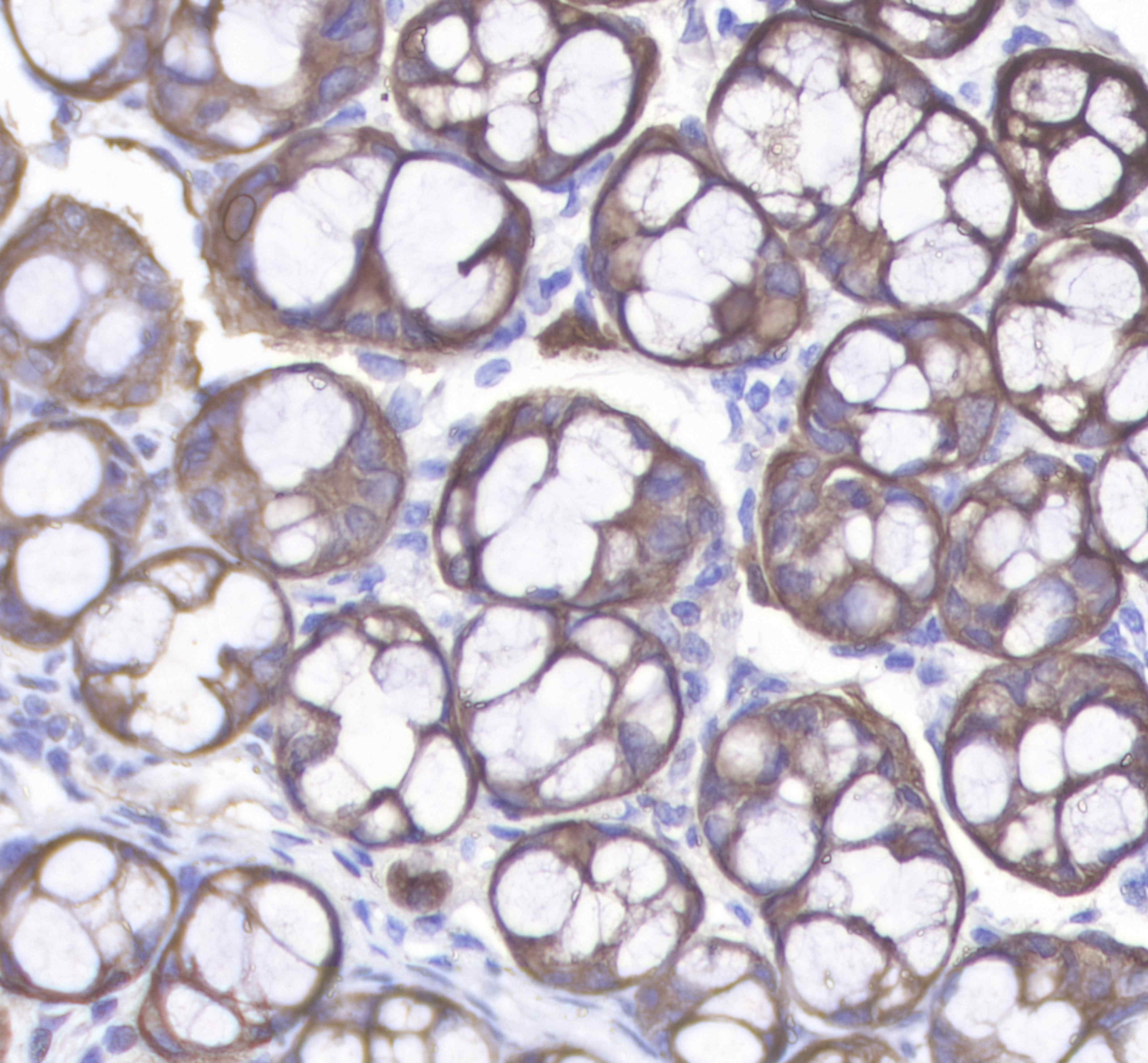
IHC shows positive staining in paraffin-embedded human kidney.
Anti-Villin antibody was used at 1/2000 dilution, followed by a Goat Anti-Rabbit IgG H&L (HRP) ready to use.
Counterstained with hematoxylin.
Heat mediated antigen retrieval with Tris/EDTA buffer pH9.0 was performed before commencing with IHC staining protocol.
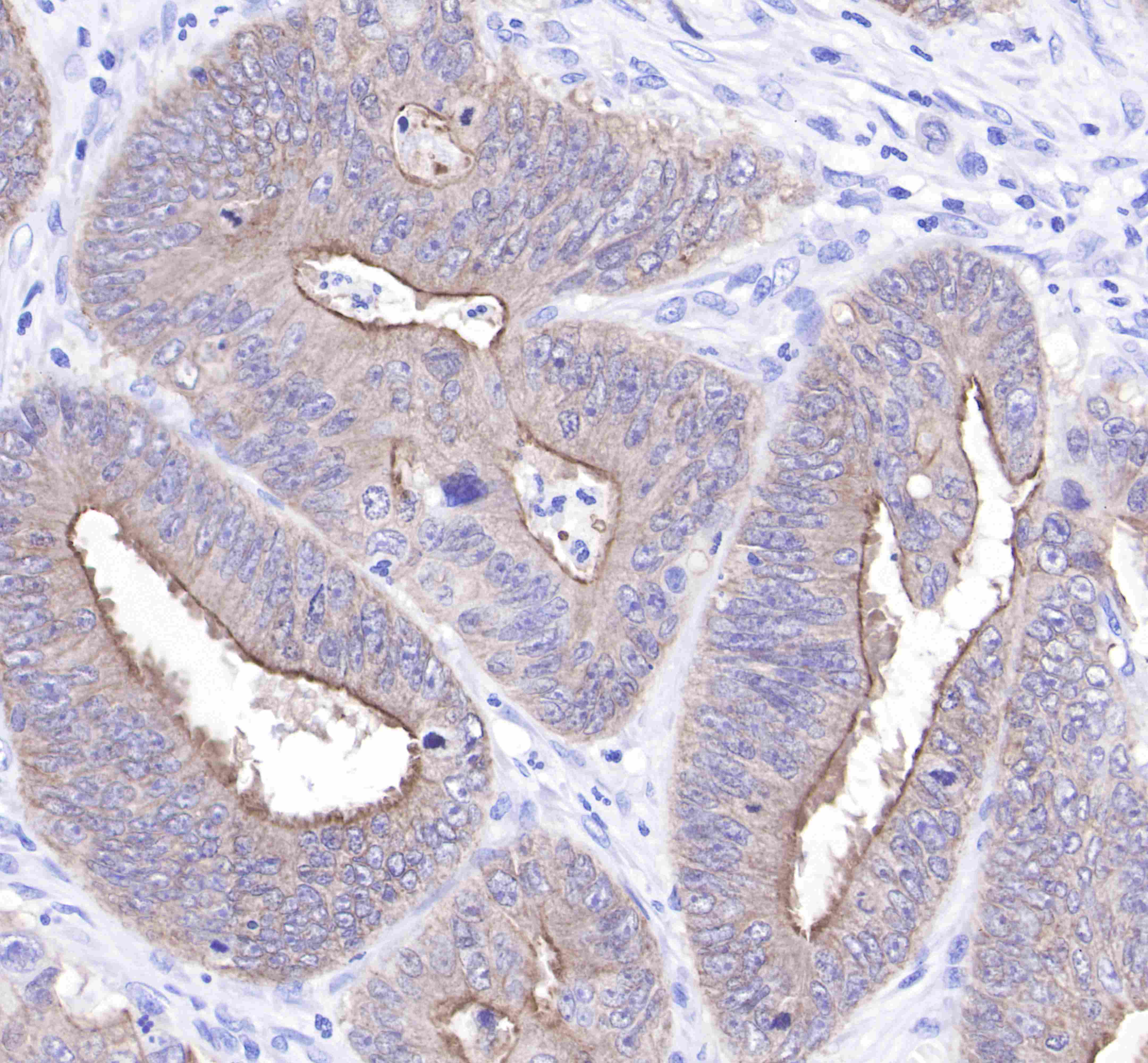
IHC shows positive staining in paraffin-embedded human colon cancer.
Anti-Villin antibody was used at 1/2000 dilution, followed by a Goat Anti-Rabbit IgG H&L (HRP) ready to use.
Counterstained with hematoxylin.
Heat mediated antigen retrieval with Tris/EDTA buffer pH9.0 was performed before commencing with IHC staining protocol.
IHC shows positive staining in paraffin-embedded mouse colon.
Anti-Villin antibody was used at 1/2000 dilution, followed by a Goat Anti-Rabbit IgG H&L (HRP) ready to use.
Counterstained with hematoxylin.
Heat mediated antigen retrieval with Tris/EDTA buffer pH9.0 was performed before commencing with IHC staining protocol.
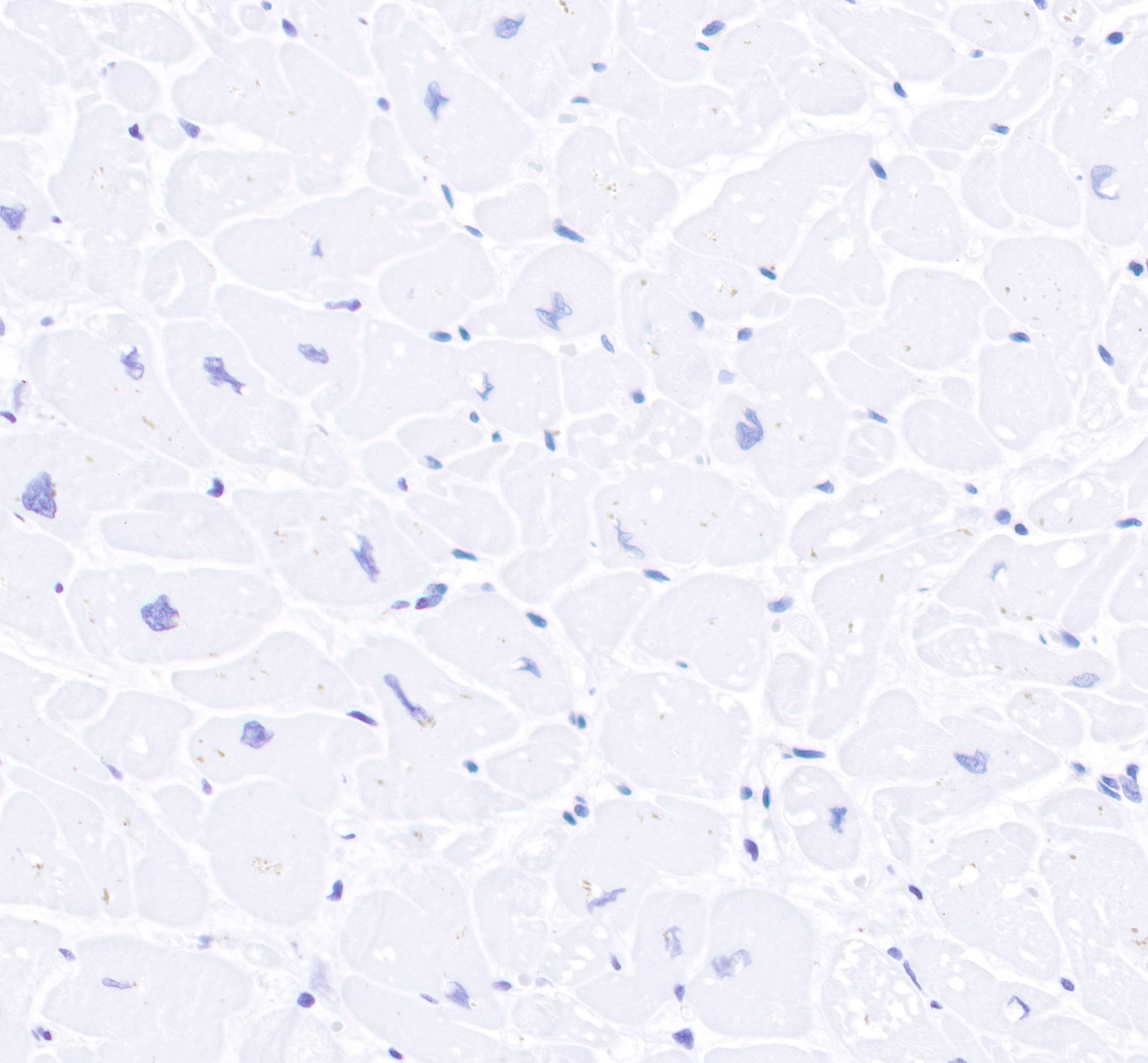
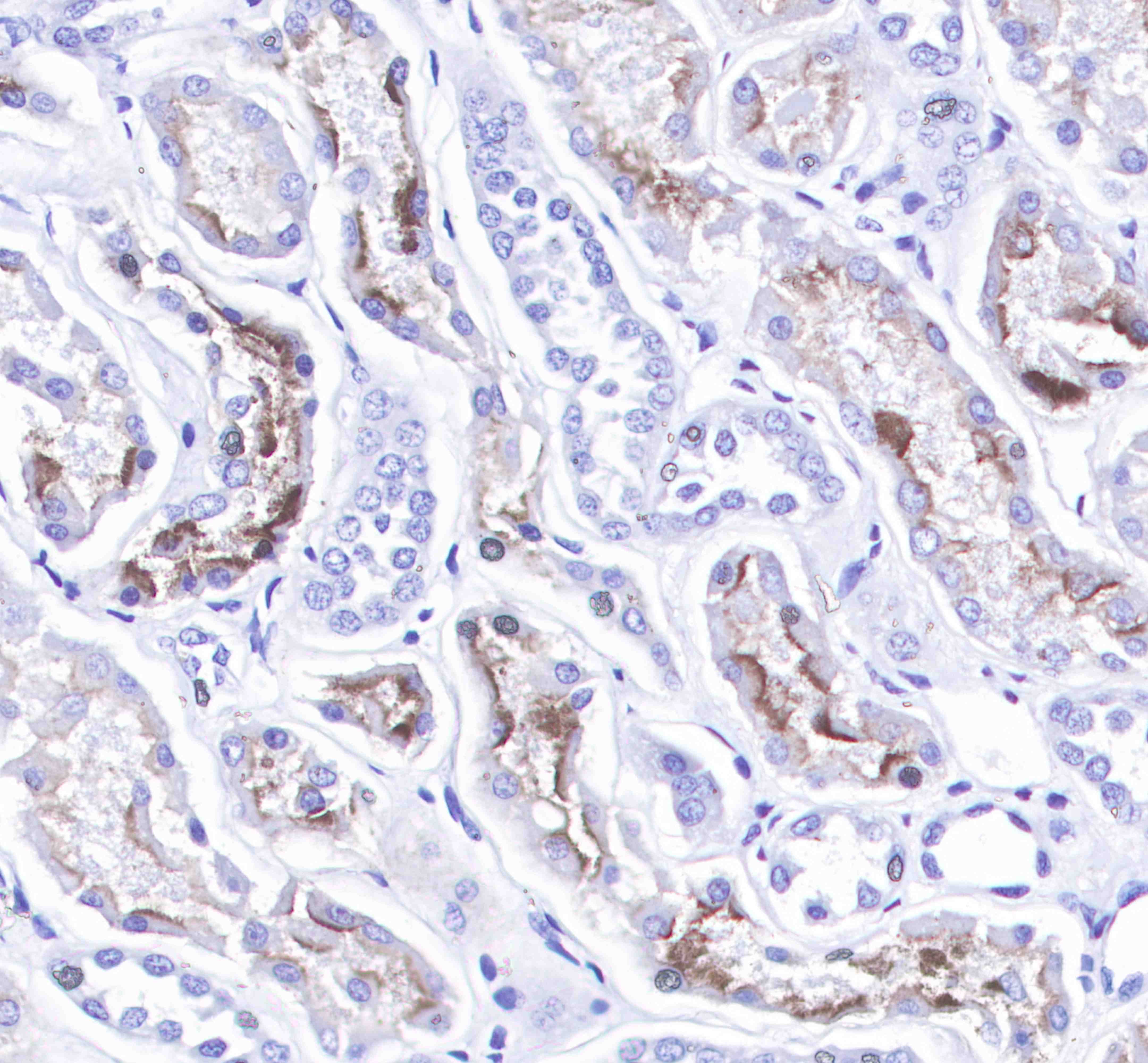
IHC shows negative staining in paraffin-embedded human myocardium(negative tissues).
Anti-Villin antibody was used at 1/2000 dilution, followed by a Goat Anti-Rabbit IgG H&L (HRP) ready to use.
Counterstained with hematoxylin.
Heat mediated antigen retrieval with Tris/EDTA buffer pH9.0 was performed before commencing with IHC staining protocol.
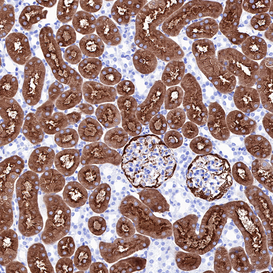
IHC shows positive staining in paraffin-embedded rat kidney. Anti-Villin antibody was used at 1/2000 dilution, followed by a Goat Anti-Rabbit IgG H&L (HRP) ready to use. Counterstained with hematoxylin. Heat mediated antigen retrieval with Tris/EDTA buffer pH9.0 was performed before commencing with IHC staining protocol.
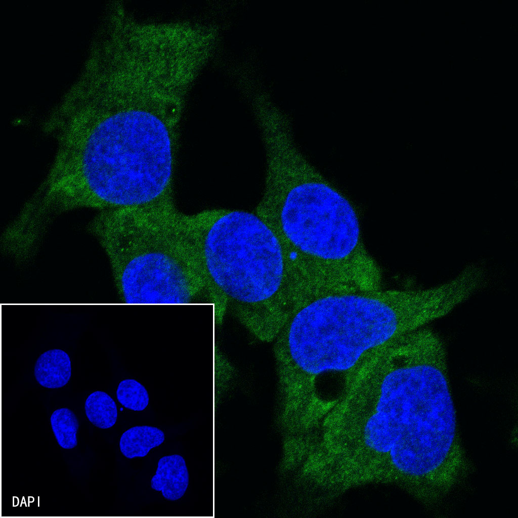
ICC shows positive staining in HepG2 cells. Anti-Villin antibody was used at 1/500 dilution (Green) and incubated overnight at 4°C. Goat polyclonal Antibody to Rabbit IgG - H&L (Alexa Fluor® 488) was used as secondary antibody at 1/1000 dilution. The cells were fixed with 4% PFA and permeabilized with 0.1% PBS-Triton X-100. Nuclei were counterstained with DAPI (Blue).
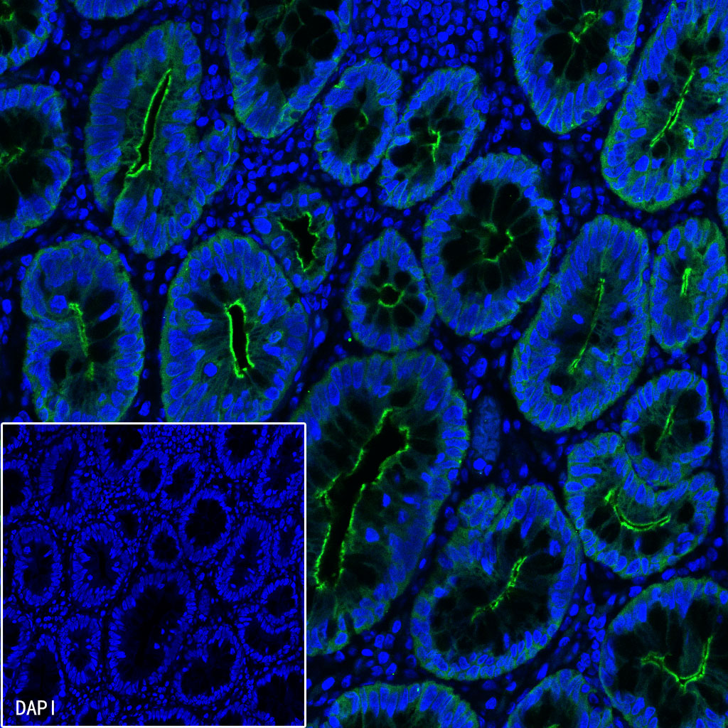
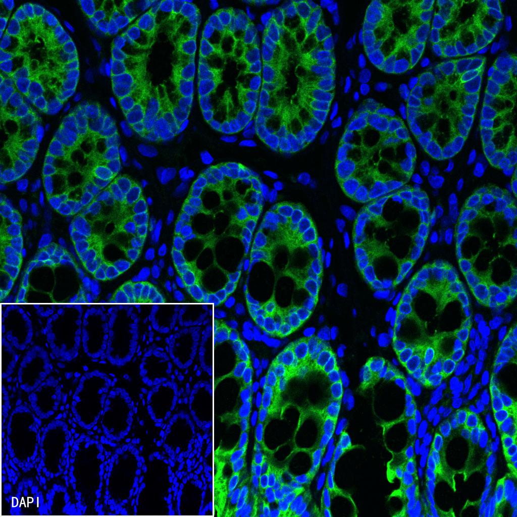
IF shows positive staining in paraffin-embedded human colon. Anti-Villin antibody was used at 1/200 dilution (Green) and incubated overnight at 4°C. Goat polyclonal Antibody to Rabbit IgG - H&L (Alexa Fluor® 488) was used as secondary antibody at 1/1000 dilution. Counterstained with DAPI (Blue). Heat mediated antigen retrieval with EDTA buffer pH9.0 was performed before commencing with IF staining protocol.
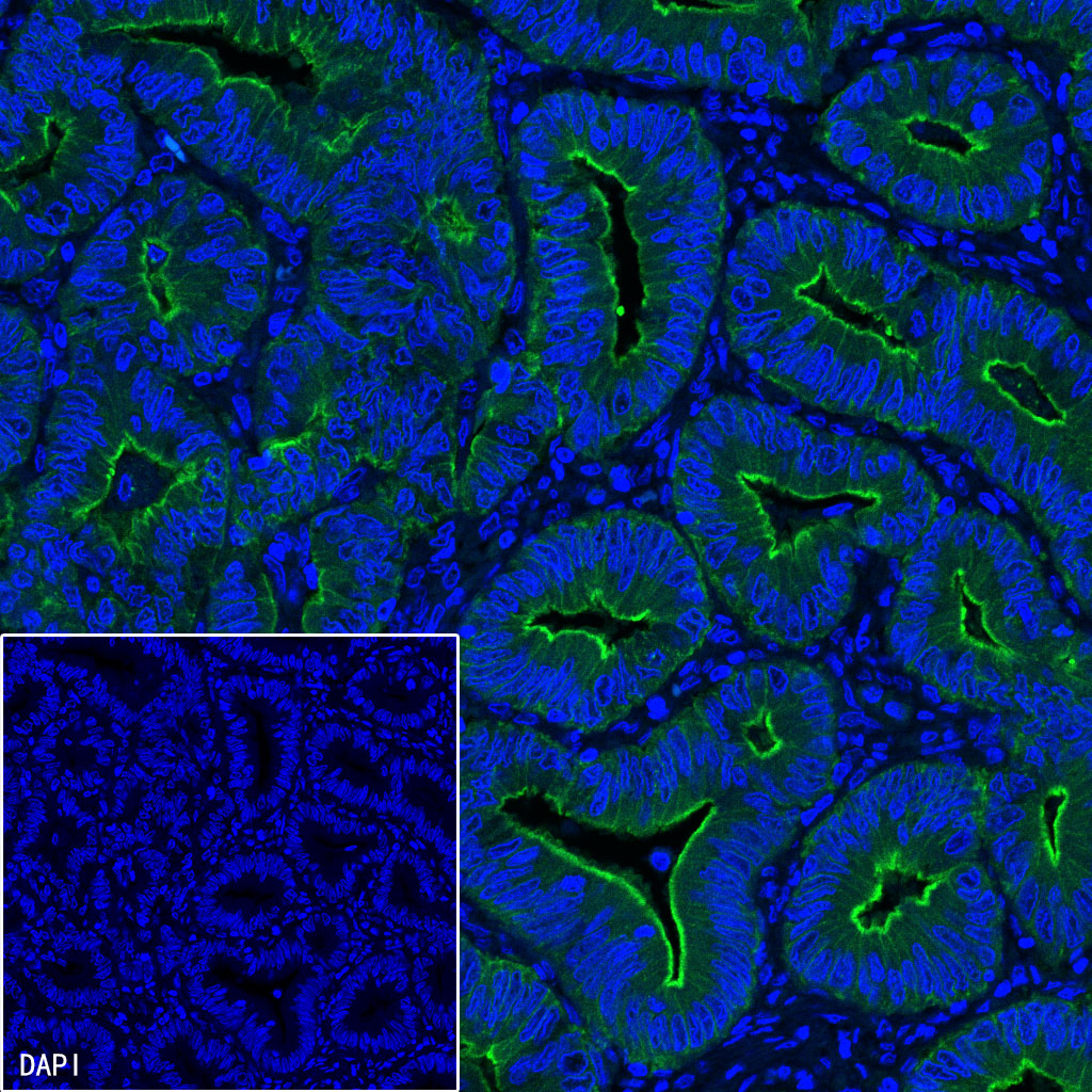
IF shows positive staining in paraffin-embedded human colon cancer. Anti-Villin antibody was used at 1/200 dilution (Green) and incubated overnight at 4°C. Goat polyclonal Antibody to Rabbit IgG - H&L (Alexa Fluor® 488) was used as secondary antibody at 1/1000 dilution. Counterstained with DAPI (Blue). Heat mediated antigen retrieval with EDTA buffer pH9.0 was performed before commencing with IF staining protocol.
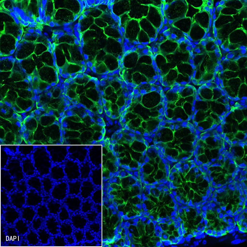
IF shows positive staining in paraffin-embedded mouse colon. Anti-Villin antibody was used at 1/1000 dilution (Green) and incubated overnight at 4°C. Goat polyclonal Antibody to Rabbit IgG - H&L (Alexa Fluor® 488) was used as secondary antibody at 1/1000 dilution. Counterstained with DAPI (Blue). Heat mediated antigen retrieval with EDTA buffer pH9.0 was performed before commencing with IF staining protocol.
IF shows positive staining in paraffin-embedded rat colon. Anti-Villin antibody was used at 1/1000 dilution (Green) and incubated overnight at 4°C. Goat polyclonal Antibody to Rabbit IgG - H&L (Alexa Fluor® 488) was used as secondary antibody at 1/1000 dilution. Counterstained with DAPI (Blue). Heat mediated antigen retrieval with EDTA buffer pH9.0 was performed before commencing with IF staining protocol.
 您现在的位置: