

12 months from date of receipt / reconstitution, -20 °C as supplied
| 应用 | 稀释度 |
|---|---|
| WB | 1:1000 |
| ICC | 1:250 |
| IHC-P | 1:250-1:1000 |
| ICFCM | 1:500 |
Keratin, type II cytoskeletal 8 also known as cytokeratin-8 (CK-8) or keratin-8 (K8) is a keratin protein that is encoded in human by the KRT8 gene. In normal tissue, it reacts mainly with secretory epithelia, but not with squamous epithelium, such as that found in the skin, cervix, and esophagus. However, it also reacts with a range of malignant cells, including those derived from secretory epithelia, but also some squamous carcinomata, such as spindle cell carcinoma. It also reacts with neuroendocrine tumors.Keratin 8 is often used together with keratin 18 and keratin 19 to differentiate cells of epithelial origin from hematopoietic cells in tests that enumerate circulating tumor cells in blood.
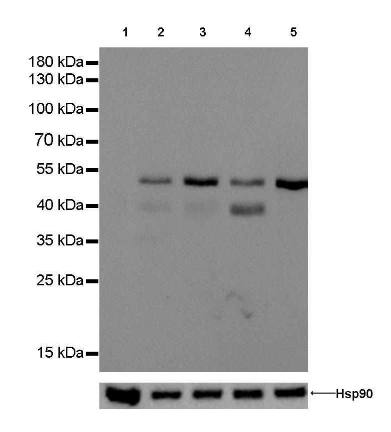
WB result of anti-Keratin 8 antibody
Primary antibody : Anti-Keratin 8 antibody at 1/1000 dilution
Lane 1 : Jurkat whole cell lysate 20 µg
Lane 2 : A549 whole cell lysate 20 µg
Lane 3 : SK-BR-3 whole cell lysate 20 µg
Lane 4 : MCF-7 whole cell lysate 20 µg
Lane 5 : Hela whole cell lysate 20 µg
Negative control: Jurkat whole cell lysate
Secondary antibody: Goat Anti-Rabbit IgG, (H+L), HRP conjugated at 1/10000 dilution
Predicted MW: 55 kDa
Observed MW: 53 kDa
Exposure time: 120 seconds
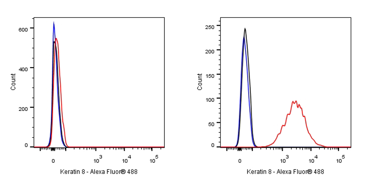
Flow cytometric analysis of 4% PFA fixed 90% methanol permeabilized Jurkat (Human T cell leukemia T lymphocyte, left) / HeLa (Human cervix adenocarcinoma epithelial cell, right) cells labelling Keratin 8 antibody at 1/500 dilution (0.1 μg) / (red) compared with a Rabbit monoclonal IgG (Black) isotype control and an unlabelled control (cells without incubation with primary antibody and secondary antibody) (Blue). Goat Anti - Rabbit IgG Alexa Fluor® 488 was used as the secondary antibody.
Negative control: Jurkat
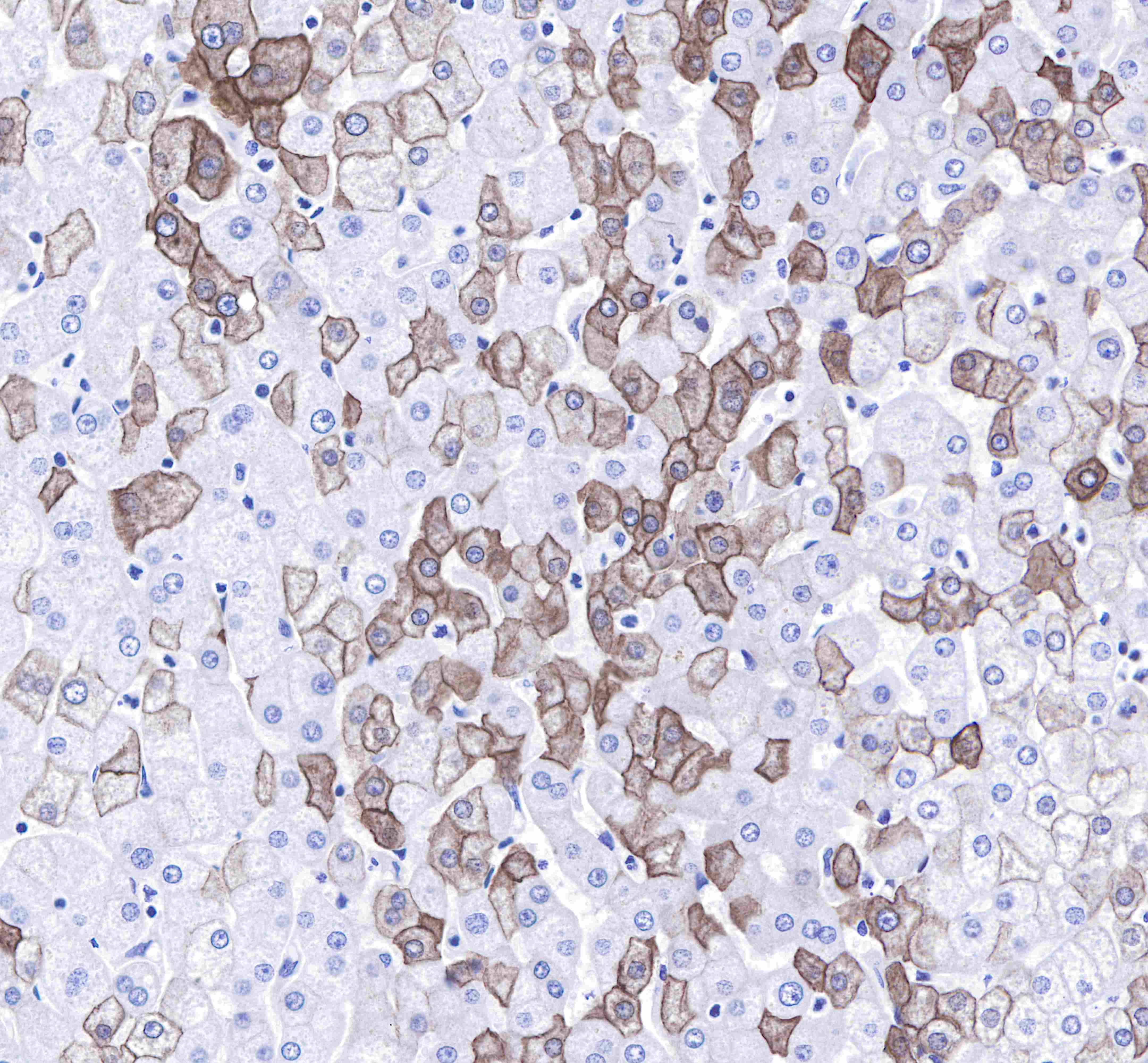
IHC shows cytoplasm and membrane staining in paraffin-embedded human liver.
Anti-Keratin 8 antibody was used at 1/250 dilution, followed by a Goat Anti-Rabbit IgG H&L (HRP) ready to use.
Counterstained with hematoxylin.
Heat mediated antigen retrieval with Tris/EDTA buffer pH9.0 was performed before commencing with IHC staining protocol.
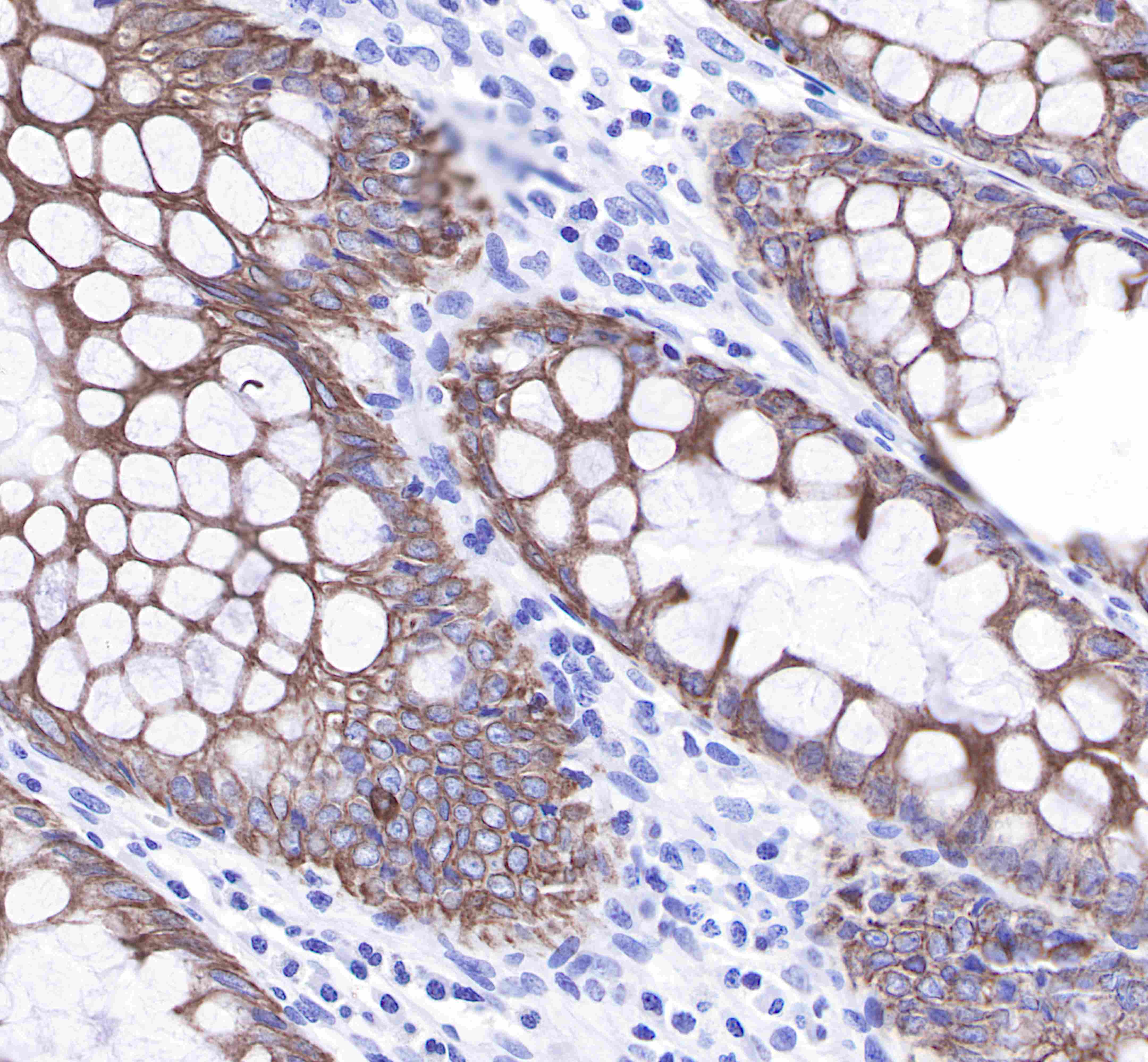
IHC shows cytoplasm and membrane staining in paraffin-embedded human colon.
Anti-Keratin 8 antibody was used at 1/250 dilution, followed by a Goat Anti-Rabbit IgG H&L (HRP) ready to use.
Counterstained with hematoxylin.
Heat mediated antigen retrieval with Tris/EDTA buffer pH9.0 was performed before commencing with IHC staining protocol.
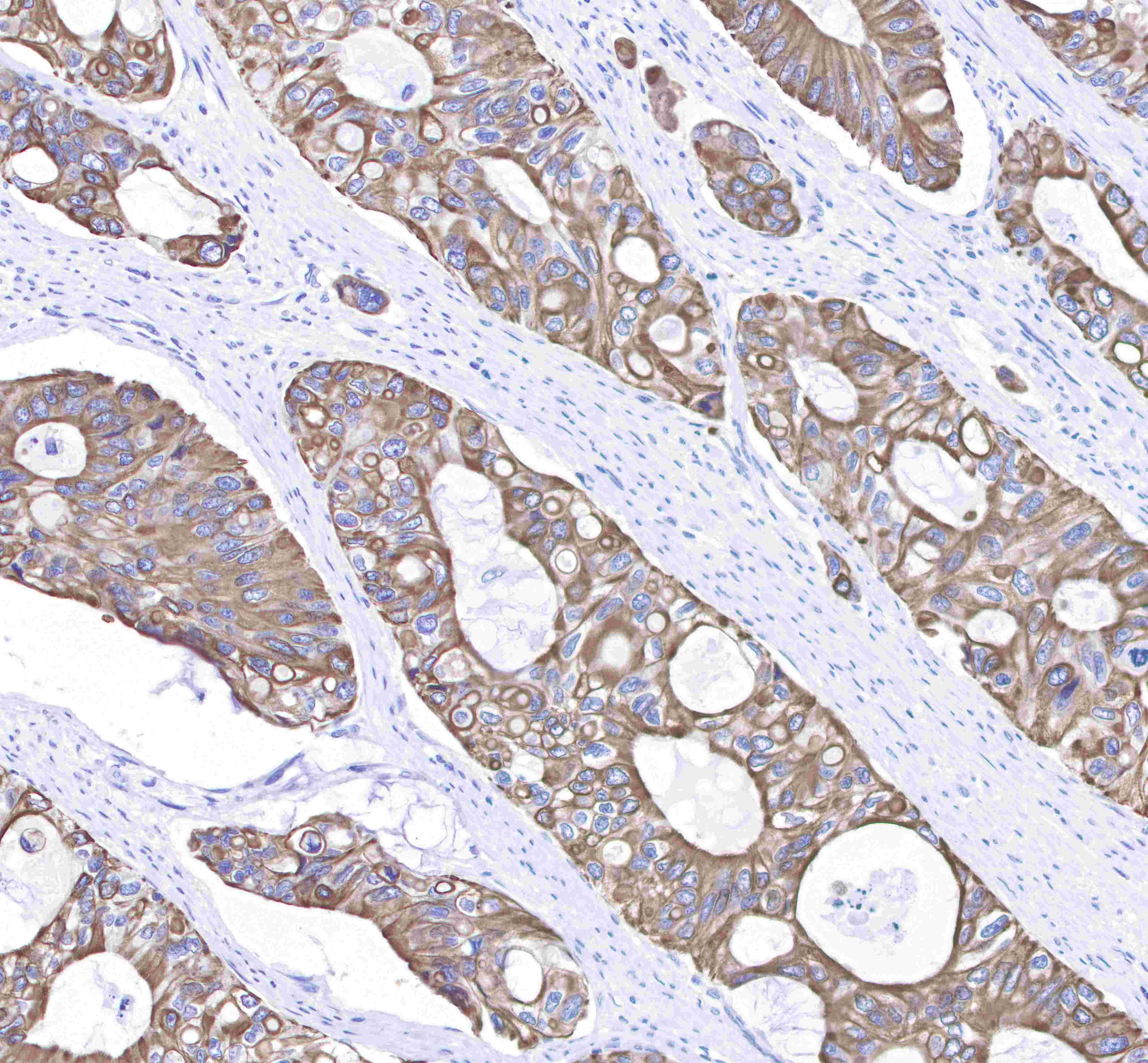
IHC shows cytoplasm and membrane staining in paraffin-embedded human lung squamous cell cancer.
Anti-Keratin 8 antibody was used at 1/250 dilution, followed by a Goat Anti-Rabbit IgG H&L (HRP) ready to use.
Counterstained with hematoxylin.
Heat mediated antigen retrieval with Tris/EDTA buffer pH9.0 was performed before commencing with IHC staining protocol.
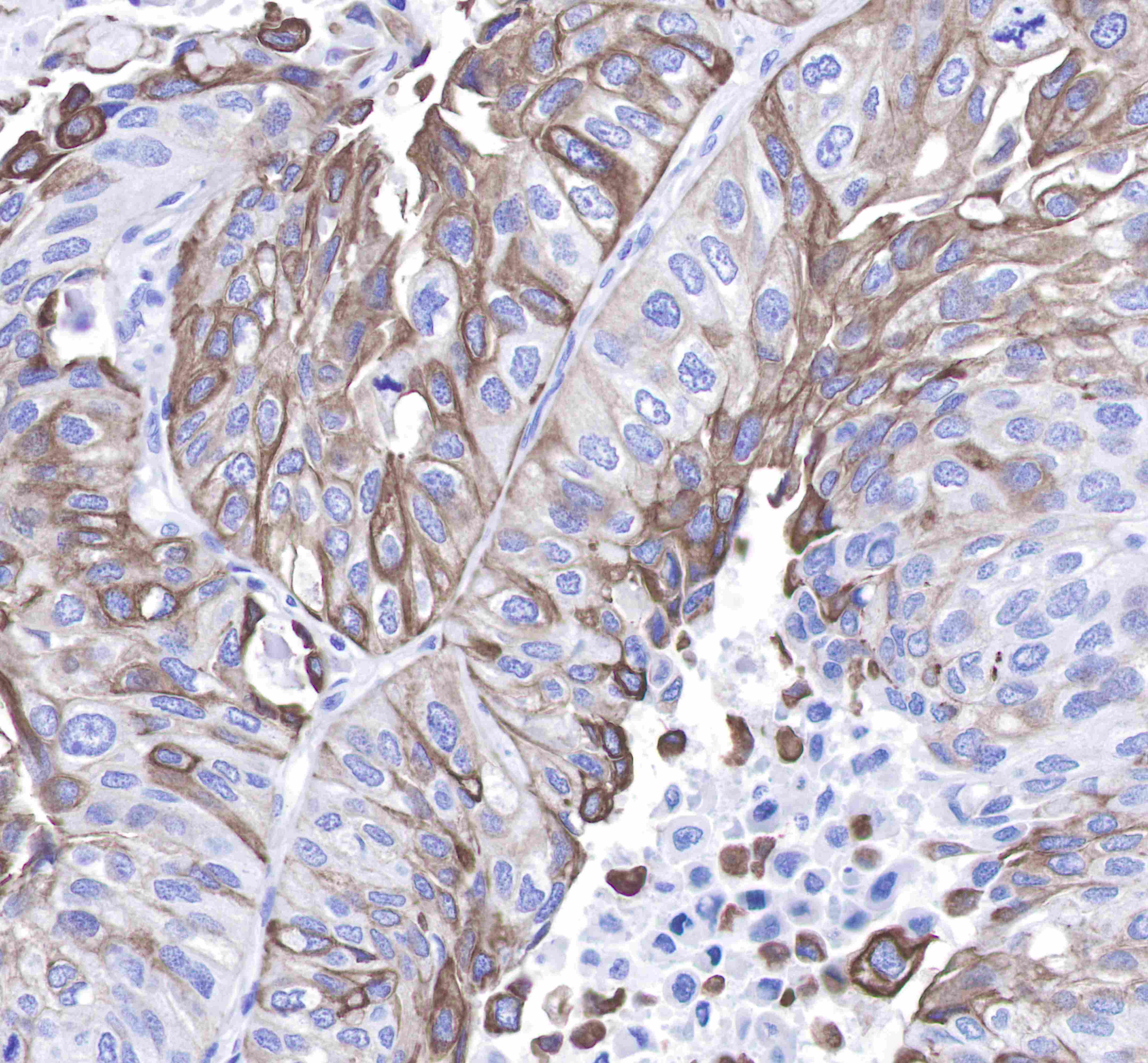
IHC shows cytoplasm and membrane staining in paraffin-embedded human colon cancer.
Anti-Keratin 8 antibody was used at 1/250 dilution, followed by a Goat Anti-Rabbit IgG H&L (HRP) ready to use.
Counterstained with hematoxylin.
Heat mediated antigen retrieval with Tris/EDTA buffer pH9.0 was performed before commencing with IHC staining protocol.
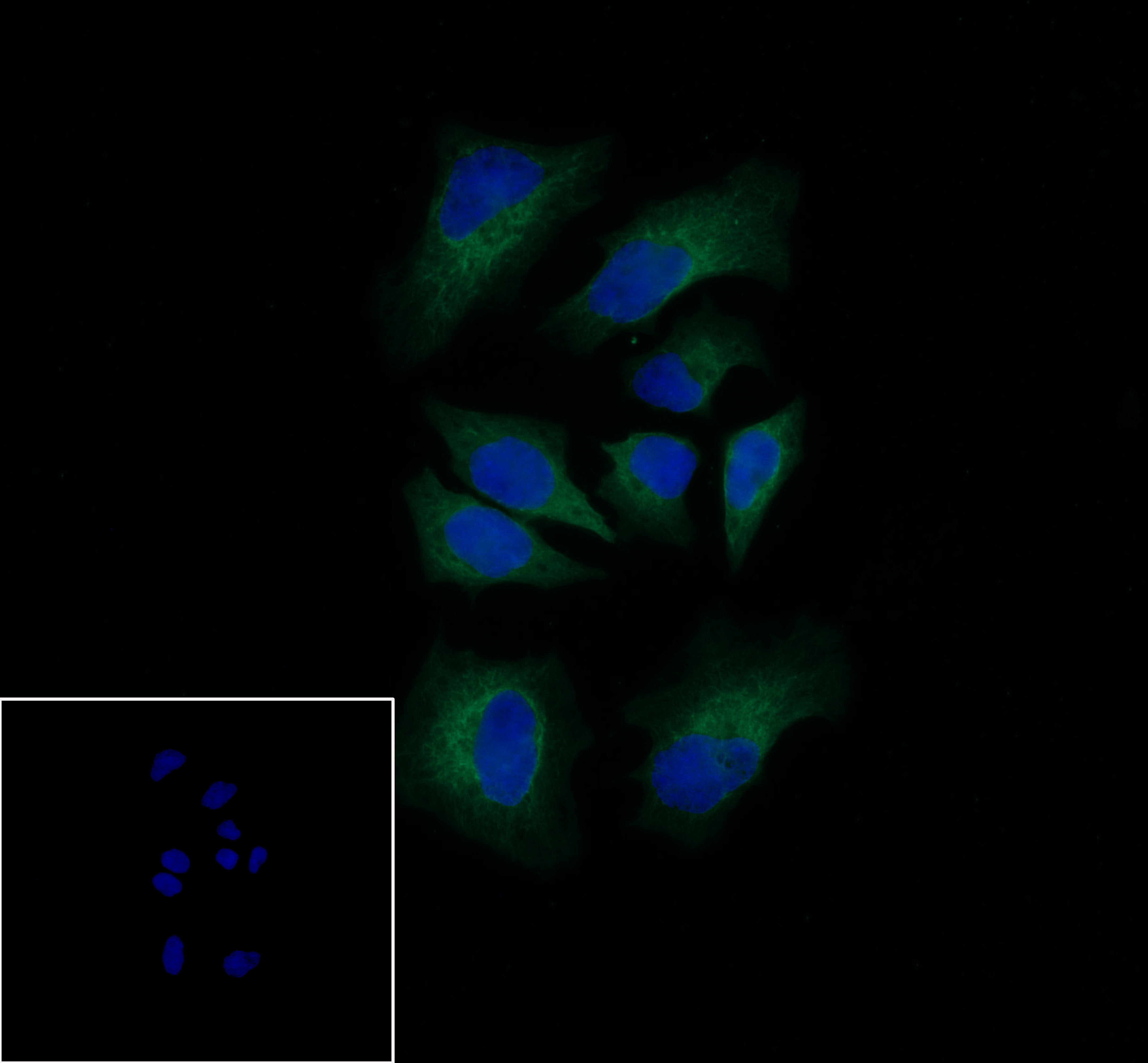
ICC shows cytoplasm staining in HeLa cells.
Anti-Keratin 8 antibody was used at 1/250 dilution and incubated overnight at 4°C.
Goat polyclonal Antibody to Rabbit IgG - H&L (Alexa Fluor® 488) was used as secondary antibody at 1/1000 dilution.
The cells were fixed with 100% methanol and permeabilized with 0.1% PBS-Triton X-100.
Nuclei were counterstained with DAPI.
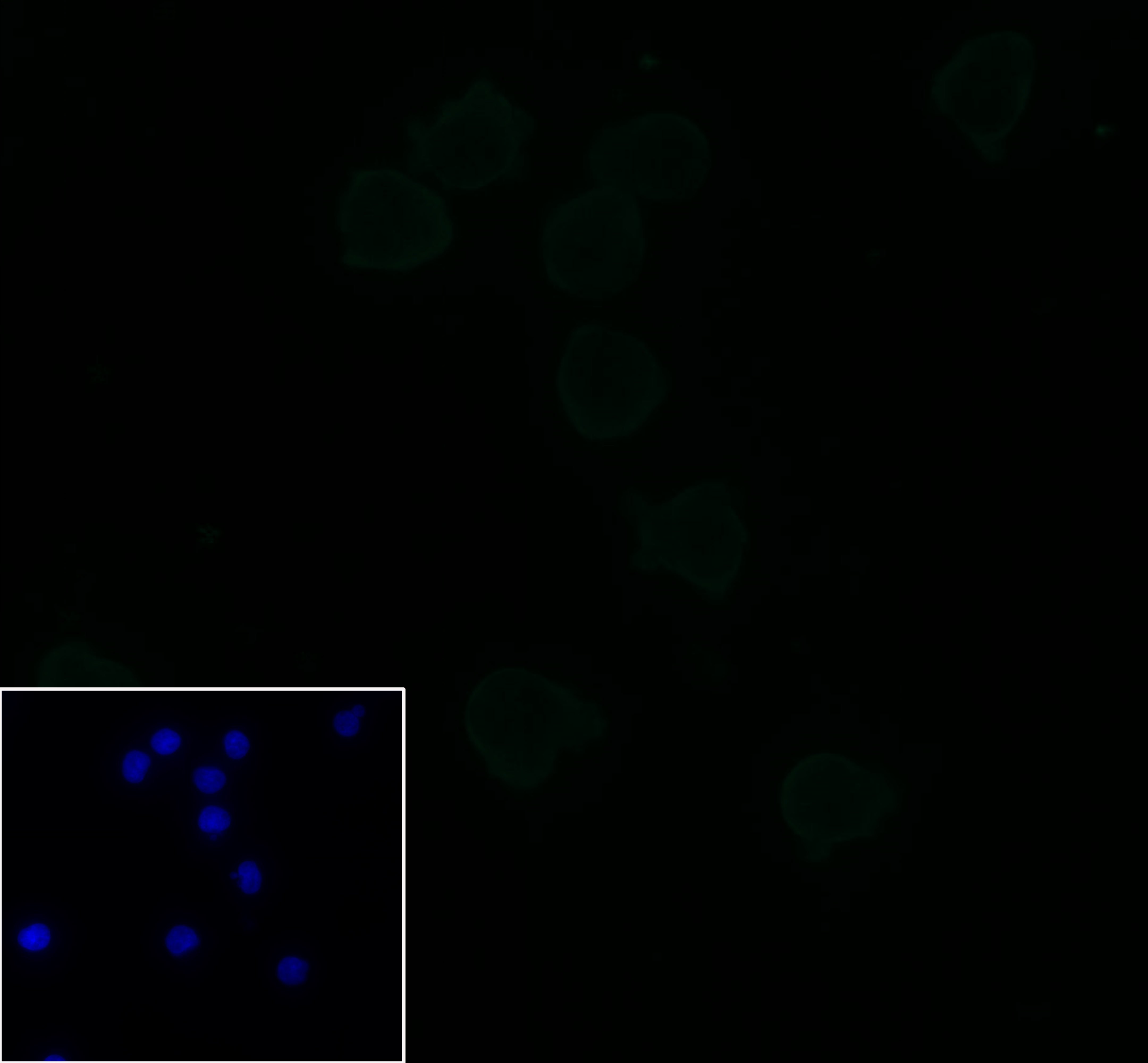
ICC shows negative staining in Jurkat cells (negative control).
Anti-Keratin 8 antibody was used at 1/250 dilution and incubated overnight at 4°C.
Goat polyclonal Antibody to Rabbit IgG - H&L (Alexa Fluor® 488) was used as secondary antibody at 1/1000 dilution.
The cells were fixed with 100% methanol and permeabilized with 0.1% PBS-Triton X-100.
Nuclei were counterstained with DAPI.
 您现在的位置: