

102kDa
PBS, 40% Glycerol, 0.05%BSA, 0.03% Proclin 300
12 months from date of receipt / reconstitution, -20 °C as supplied
| 应用 | 稀释度 |
|---|---|
| IHC-P | 1:1000 |
| ICC | 1:100 |
| WB | 1:1000 |
| ICFCM | 1:500 |
| IF | 1:500 |
MCM2 is one of the highly conserved mini-chromosome maintenance proteins (MCM) that are involved in the initiation of eukaryotic genome replication. The hexameric protein complex formed by MCM proteins is a key component of the pre-replication complex (pre-RC) and may be involved in the formation of replication forks and in the recruitment of other DNA replication related proteins. MCM2 forms a complex with MCM4, 6, and 7, and has been shown to regulate the helicase activity of the complex. MCM2 is phosphorylated, and thus regulated by protein kinases CDC2 and CDC7.
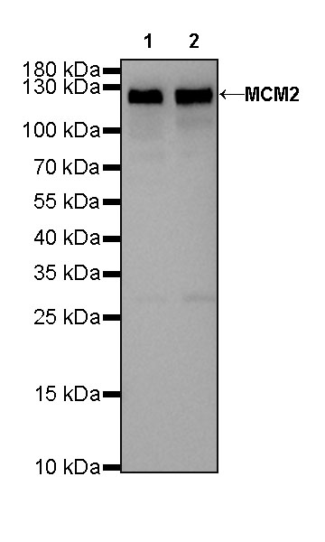
WB result of MCM2 Rabbit mAb
Primary antibody : MCM2 Rabbit mAb at 1/1000 dilution
Lane 1 : Hela whole cell lysate 20 µg
Lane 2 : Ramos whole cell lysate 20 µg
Secondary antibody: Goat Anti-Rabbit IgG, (H+L), HRP conjugated at 1/10000 dilution
Predicted MW: 125 kDa
Observed MW: 125 kDa
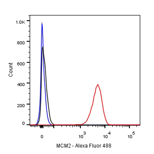
Flow cytometric analysis of Jurkat cells labelling MCM2 antibody at 1/500 (0.1 μg) dilution/ (red) compared with a Rabbit monoclonal IgG (Black) isotype control and an unlabelled control (cells without incubation with primary antibody and secondary antibody) (Blue). Goat Anti-Rabbit IgG Alexa Fluor® 488 was used as the secondary antibody.
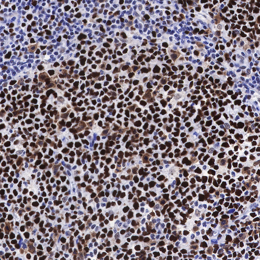
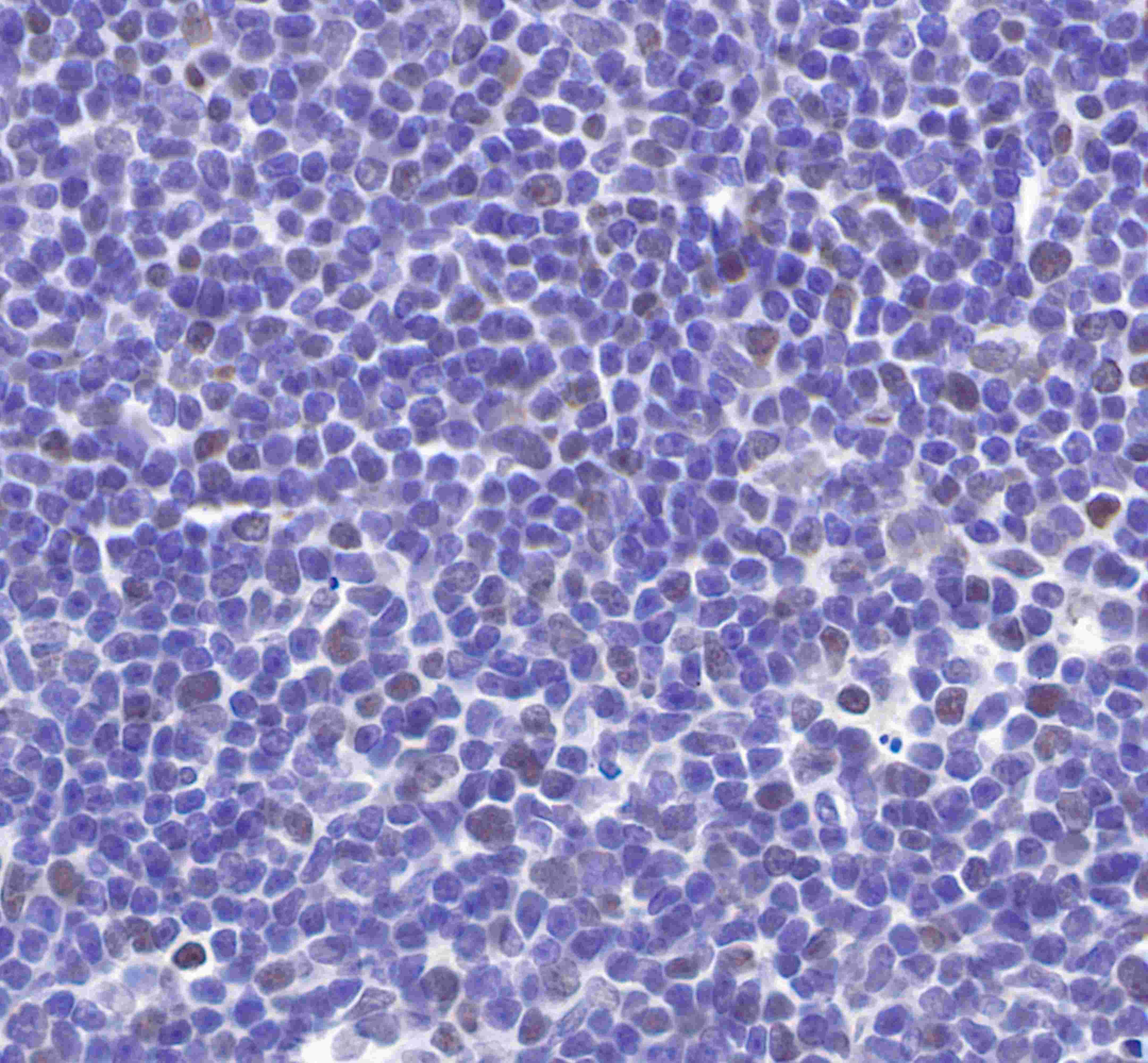
IHC show nucleus staining in paraffin-embedded human tonsil. Anti-MCM2 antibody was used at 1/1000 dilution, followed by a Goat Anti-Rabbit IgG H&L (HRP) ready to use. Counterstained with hematoxylin.
Heat mediated antigen retrieval with Tris/EDTA buffer pH9.0 was performed before commencing with IHC staining protocol.
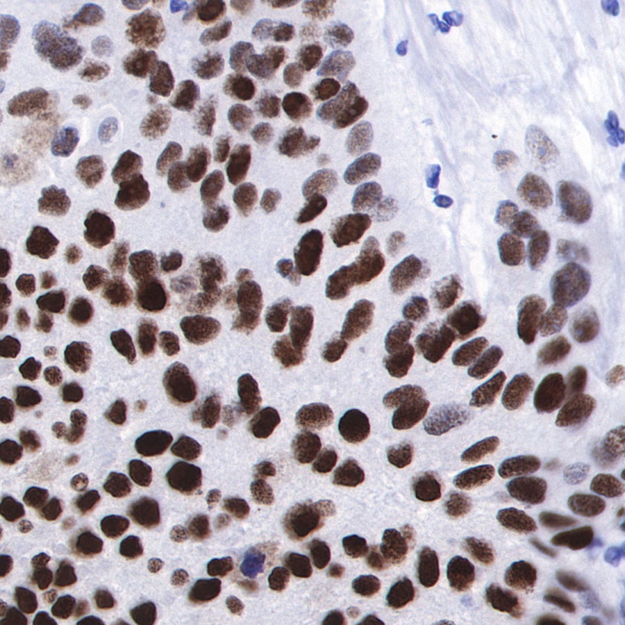
IHC shows nucleus staining in paraffin-embedded human bladder cancer. Anti-MCM2 antibody was used at 1/1000 dilution, followed by a Goat Anti-Rabbit IgG H&L (HRP) ready to use. Counterstained with hematoxylin.
Heat mediated antigen retrieval with Tris/EDTA buffer pH9.0 was performed before commencing with IHC staining protocol.
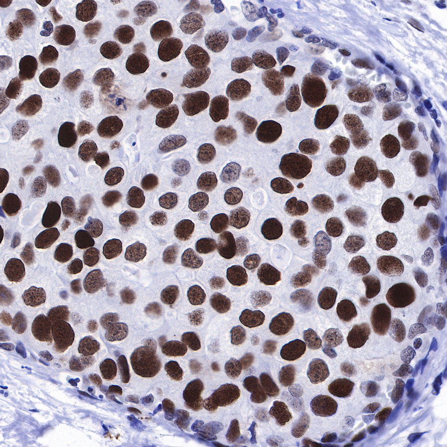
IHC shows nucleus staining in paraffin-embedded human breast cancer. Anti-MCM2 antibody was used at 1/1000 dilution, followed by a Goat Anti-Rabbit IgG H&L (HRP) ready to use. Counterstained with hematoxylin.
Heat mediated antigen retrieval with Tris/EDTA buffer pH9.0 was performed before commencing with IHC staining protocol.

IHC shows nucleus staining in paraffin-embedded human lung adenocarcinoma. Anti-MCM2 antibody was used at 1/1000 dilution, followed by a Goat Anti-Rabbit IgG H&L (HRP) ready to use. Counterstained with hematoxylin.
Heat mediated antigen retrieval with Tris/EDTA buffer pH9.0 was performed before commencing with IHC staining protocol.
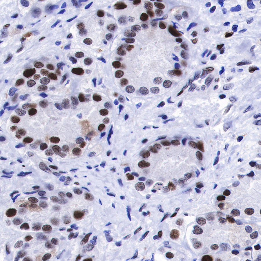
IHC shows nucleus staining in paraffin-embedded human pancreatic carcinoma. Anti-MCM2 antibody was used at 1/1000 dilution, followed by a Goat Anti-Rabbit IgG H&L (HRP) ready to use. Counterstained with hematoxylin.
Heat mediated antigen retrieval with Tris/EDTA buffer pH9.0 was performed before commencing with IHC staining protocol.
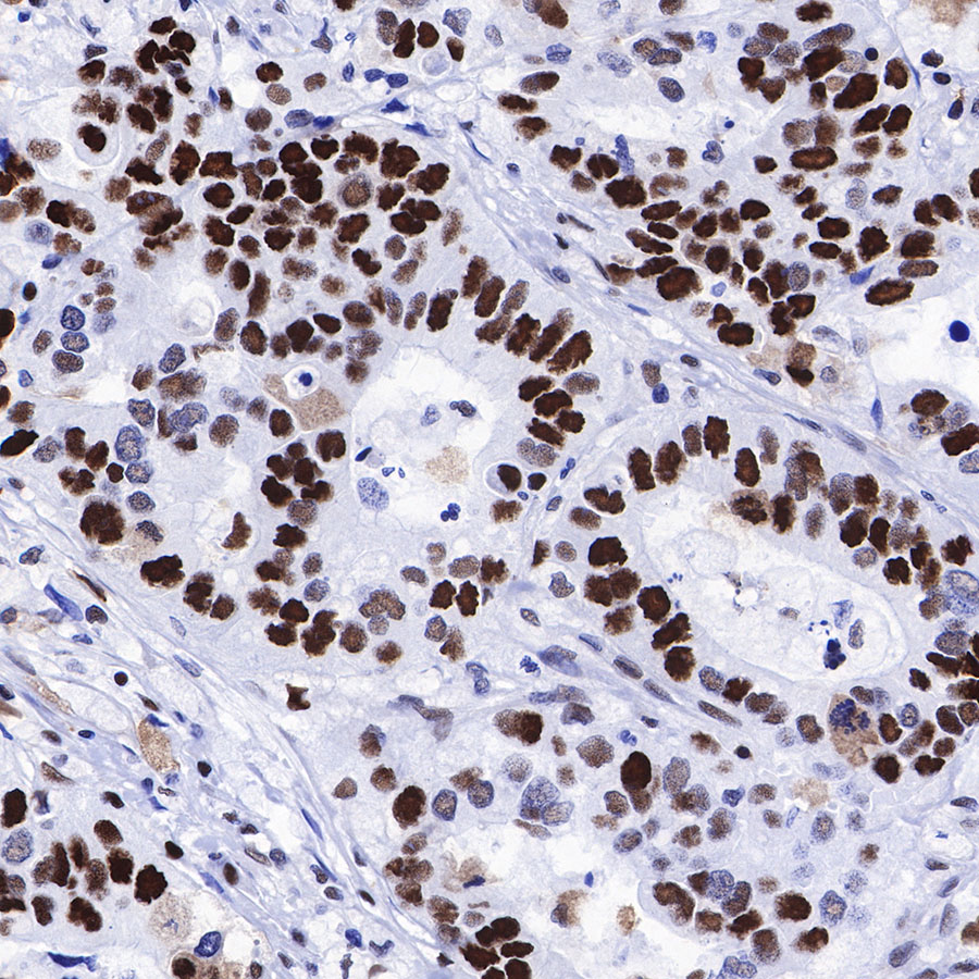
IHC shows nucleus staining in paraffin-embedded human gastric carcinoma. Anti-MCM2 antibody was used at 1/1000 dilution, followed by a Goat Anti-Rabbit IgG H&L (HRP) ready to use. Counterstained with hematoxylin.
Heat mediated antigen retrieval with Tris/EDTA buffer pH9.0 was performed before commencing with IHC staining protocol.
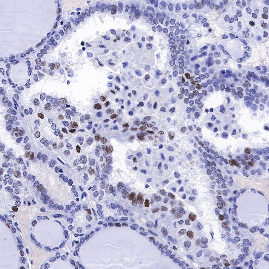
IHC shows nucleus staining in paraffin-embedded human thyroid carcinoma. Anti-MCM2 antibody was used at 1/1000 dilution, followed by a Goat Anti-Rabbit IgG H&L (HRP) ready to use. Counterstained with hematoxylin.
Heat mediated antigen retrieval with Tris/EDTA buffer pH9.0 was performed before commencing with IHC staining protocol.
IHC show nucleus staining in paraffin-embedded mouse spleen. Anti-MCM2 antibody was used at 1/1000 dilution, followed by a Goat Anti-Rabbit IgG H&L (HRP) ready to use. Counterstained with hematoxylin.
Heat mediated antigen retrieval with Tris/EDTA buffer pH9.0 was performed before commencing with IHC staining protocol.
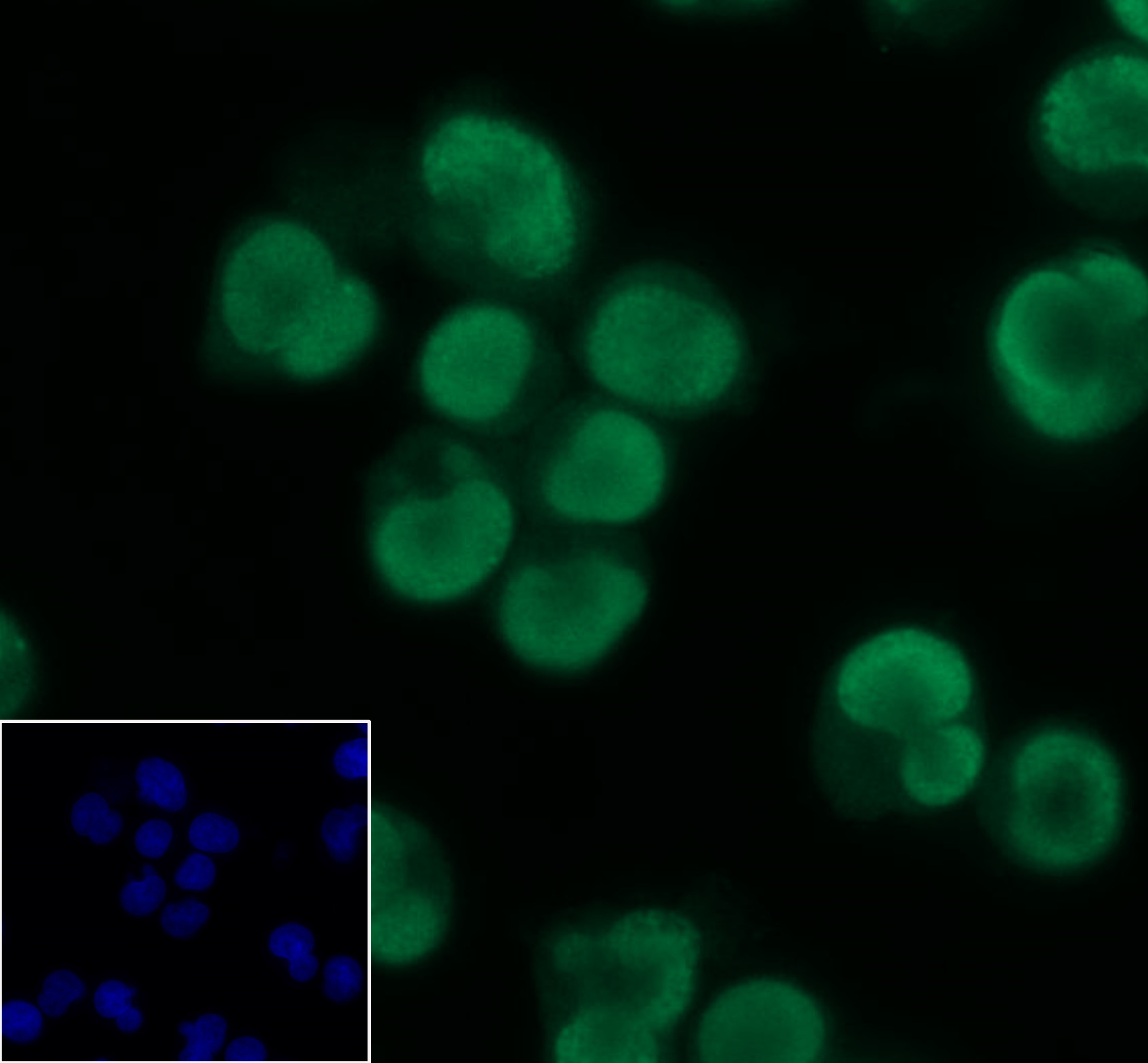
ICC shows nucleus staining in Jurkat cells. Anti-MCM2 antibody was used at 1/100 dilution and incubated overnight at 4°C. Goat polyclonal Antibody to Rabbit IgG - H&L (Alexa Fluor® 488) at 1/1000 dilution was used as secondary antibody.The cells were fixed with 4% formaldehyde and permeabilized with 0.1% PBS-Triton X-100. Nuclei were countersained with DAPI.

IF shows positive staining in paraffin-embedded human breast cancer. Anti-MCM2 antibody was used at 1/500 dilution (magenta) and incubated overnight at 4°C. Goat polyclonal Antibody to Rabbit IgG - H&L (Alexa Fluor® 647)(S0B4005) was used as secondary antibody at 1/500 dilution. Counterstained with DAPI (Blue). Heat mediated antigen retrieval with EDTA buffer pH9.0 was performed before commencing with IF staining protocol.

IF shows positive staining in paraffin-embedded human colon cancer. Anti-MCM2 antibody was used at 1/500 dilution (magenta) and incubated overnight at 4°C. Goat polyclonal Antibody to Rabbit IgG - H&L (Alexa Fluor® 647)(S0B4005) was used as secondary antibody at 1/500 dilution. Counterstained with DAPI (Blue). Heat mediated antigen retrieval with EDTA buffer pH9.0 was performed before commencing with IF staining protocol.
 您现在的位置: