

60kDa
12 months from date of receipt / reconstitution, -20 °C as supplied
| 应用 | 稀释度 |
|---|---|
| IHC-P | 1:1000 |
| WB | 1:25000 |
Keratin 6C is a type II cytokeratin, one of a number of isoforms of keratin 6 encoded by separate genes located within the type II keratin gene cluster on human chromosome 12q. Keratins are the intermediate filament proteins that form a dense meshwork of filaments throughout the cytoplasm of epithelial cells. Keratins form heteropolymers consisting of a type I and a type II keratin. Keratins are generally expressed in particular pairs of type I and type II keratin proteins in a tissue-specific and cellular differentiation-specific manner.
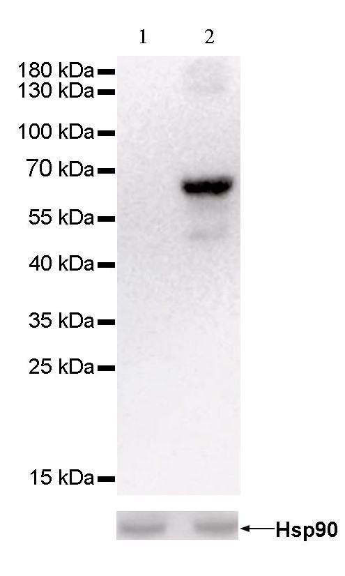
WB result of anti-Keratin 6 antibody
Primary antibody : Anti-Keratin 6 antibody at 1/25000 dilution
Lane 1 : Hela whole cell lysate 20 µg
Lane 2 : A431 whole cell lysate 20 µg
Negative control: Hela whole cell lysate
Secondary antibody: Goat Anti-Rabbit IgG, (H+L), HRP conjugated at 1/10000 dilution
Predicted MW: 60 kDa
Observed MW: 60 kDa
econds
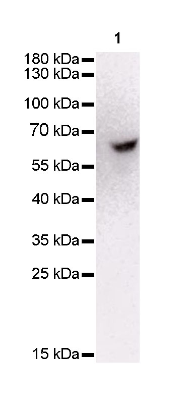
WB result of anti-Keratin 6 antibody
Primary antibody : Anti-Keratin 6 antibody at 1/25000 dilution
Lane 1 : mouse skin lysate 20 µg
Secondary antibody: Goat Anti-Rabbit IgG, (H+L), HRP conjugated at 1/10000 dilution
Predicted MW: 60 kDa
Observed MW: 60 kDa
econds
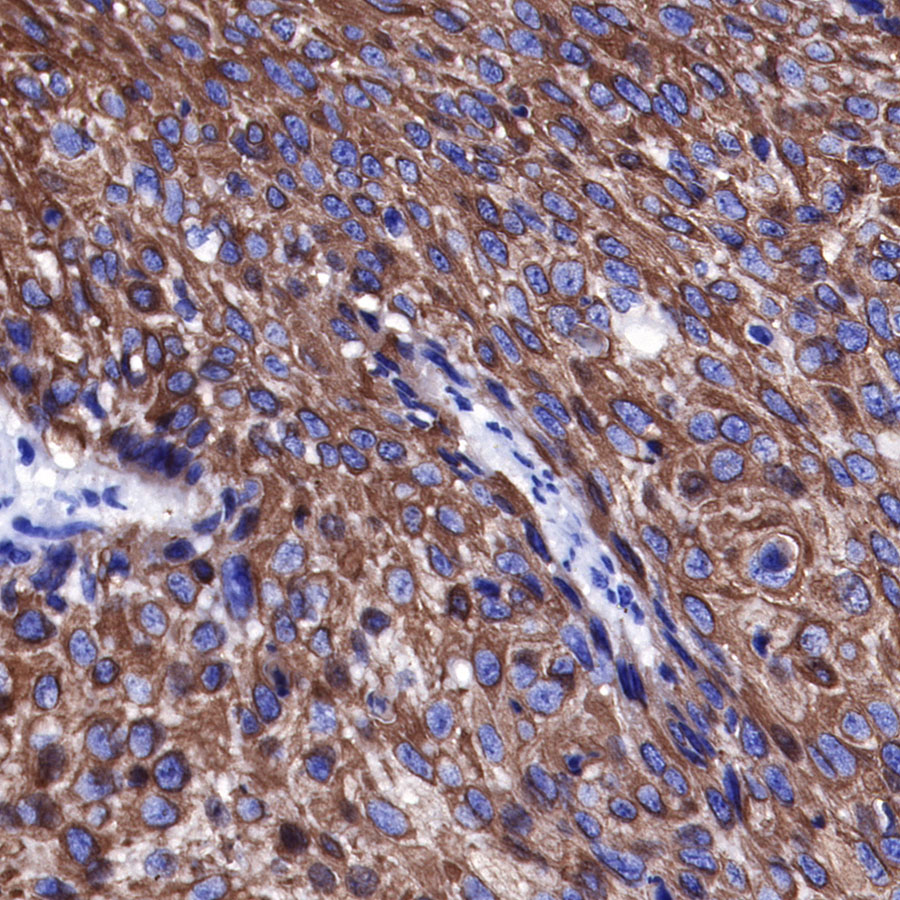
IHC shows positive staining in paraffin-embedded human cervical squamous cell carcinoma. Anti-Keratin 6 antibody was used at 1/1000 dilution, followed by a Goat Anti-Rabbit IgG H&L (HRP) ready to use. Counterstained with hematoxylin. Heat mediated antigen retrieval with Tris/EDTA buffer pH9.0 was performed before commencing with IHC staining protocol.
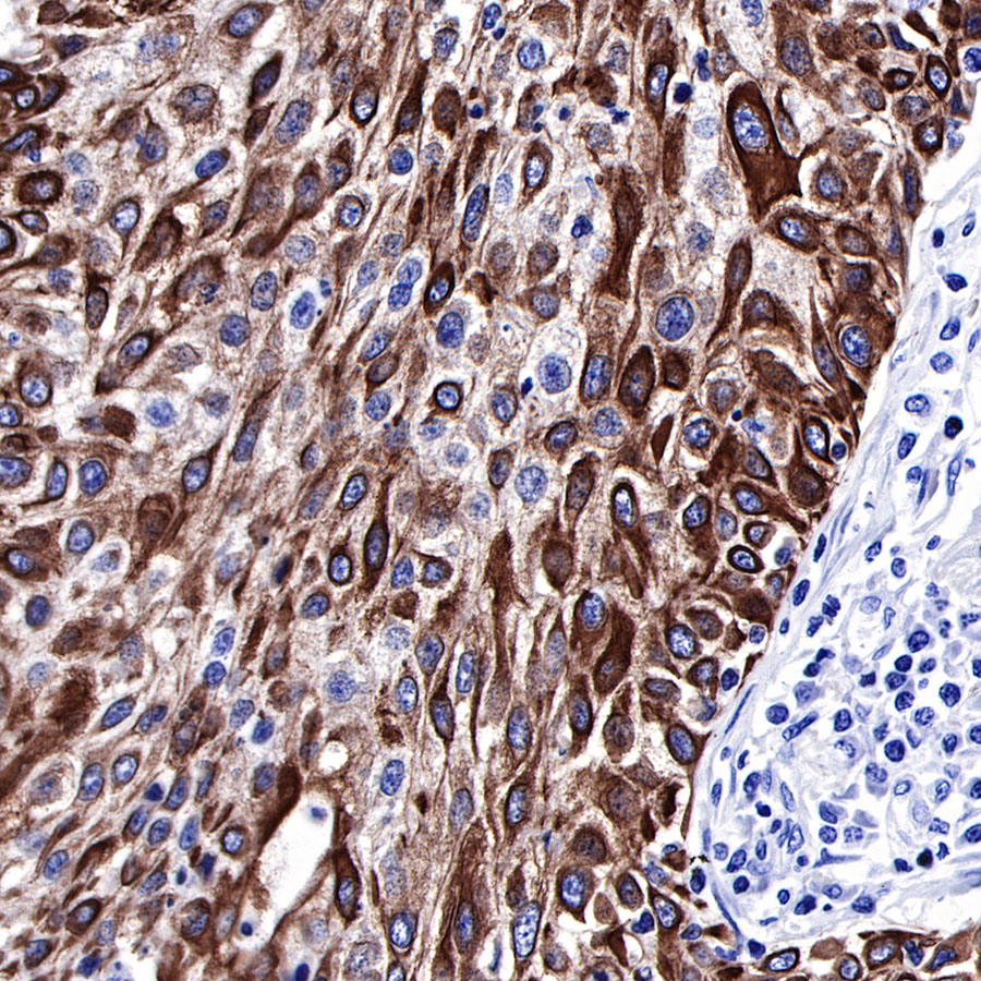
IHC shows positive staining in paraffin-embedded human lung squamous cell carcinoma. Anti-Keratin 6 antibody was used at 1/1000 dilution, followed by a Goat Anti-Rabbit IgG H&L (HRP) ready to use. Counterstained with hematoxylin. Heat mediated antigen retrieval with Tris/EDTA buffer pH9.0 was performed before commencing with IHC staining protocol.
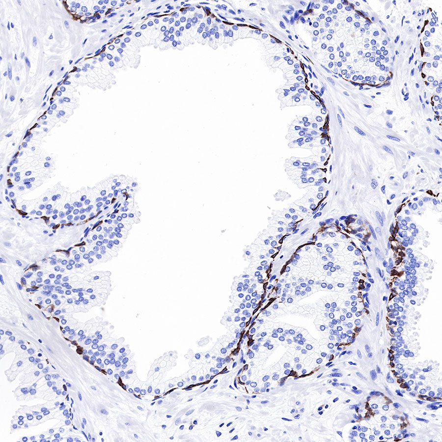
IHC shows positive staining in paraffin-embedded human prostatic hyperplasia. Anti-Keratin 6 antibody was used at 1/1000 dilution, followed by a Goat Anti-Rabbit IgG H&L (HRP) ready to use. Counterstained with hematoxylin. Heat mediated antigen retrieval with Tris/EDTA buffer pH9.0 was performed before commencing with IHC staining protocol.
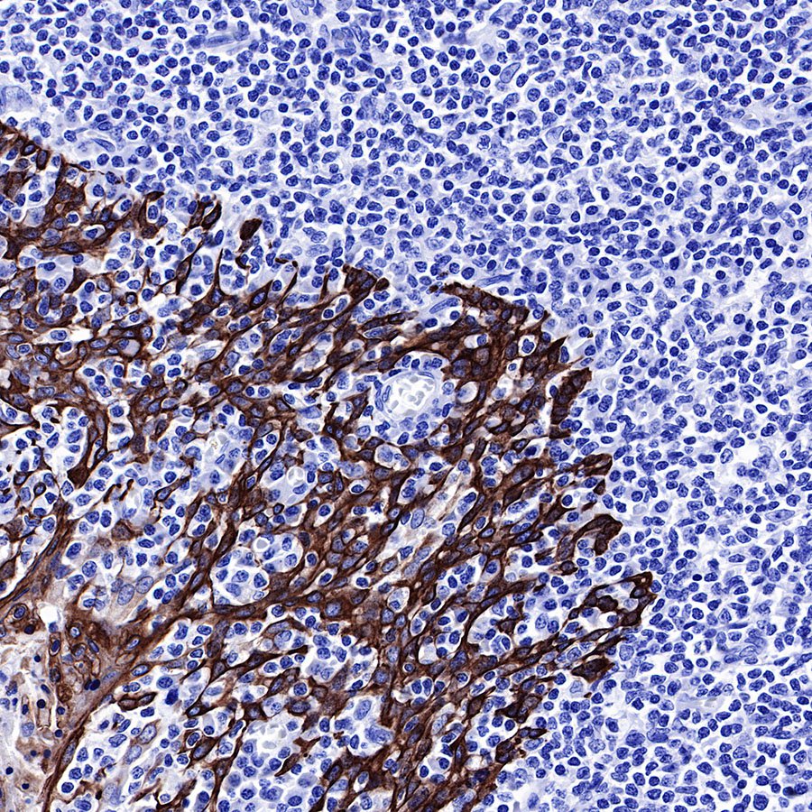
IHC shows membrane staining in paraffin-embedded human tonsil. Anti-Keratin 6 antibody was used at 1/1000 dilution, followed by a Goat Anti-Rabbit IgG H&L (HRP) ready to use. Counterstained with hematoxylin. Heat mediated antigen retrieval with Tris/EDTA buffer pH9.0 was performed before commencing with IHC staining protocol.
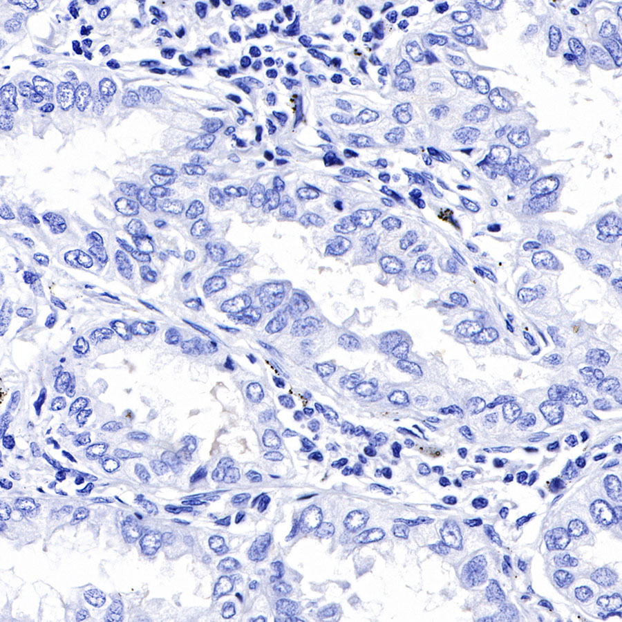
Negative control: IHC shows negative staining in paraffin-embedded human lung adenocarcinoma. Anti-Keratin 6 antibody was used at 1/1000 dilution, followed by a Goat Anti-Rabbit IgG H&L (HRP) ready to use. Counterstained with hematoxylin. Heat mediated antigen retrieval with Tris/EDTA buffer pH9.0 was performed before commencing with IHC staining protocol.
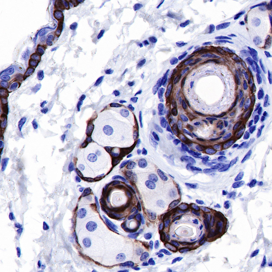
IHC shows membrane staining in paraffin-embedded mouse skin. Anti-Keratin 6 antibody was used at 1/1000 dilution, followed by a Goat Anti-Rabbit IgG H&L (HRP) ready to use. Counterstained with hematoxylin. Heat mediated antigen retrieval with Tris/EDTA buffer pH9.0 was performed before commencing with IHC staining protocol.
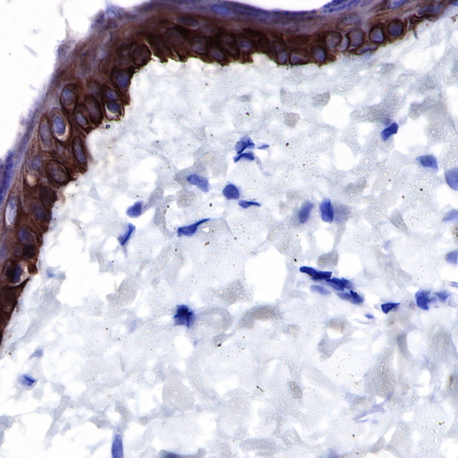
IHC shows membrane staining in paraffin-embedded rat skin. Anti-Keratin 6 antibody was used at 1/1000 dilution, followed by a Goat Anti-Rabbit IgG H&L (HRP) ready to use. Counterstained with hematoxylin. Heat mediated antigen retrieval with Tris/EDTA buffer pH9.0 was performed before commencing with IHC staining protocol.
 您现在的位置: