
12 months from date of receipt / reconstitution, -20 °C as supplied
| 应用 | 稀释度 |
|---|---|
| WB | 1:1000 |
| IHC-P | 1:100 |
B7-H3 is a 316 amino acid-long type I transmembrane protein, existing in two isoforms determined by its extracellular domain. In humans it consists of one pair (2Ig-B7-H3) or two identical pairs (4Ig-B7-H3) due to exon duplication. B7-H3 mRNA is expressed in most normal tissues. In contrast, B7-H3 protein has a very limited expression on normal tissues because of its post-transcriptional regulation by microRNAs. However, B7-H3 protein is expressed at high frequency on many different cancer types (60% of all cancers). In non-malignant tissues, B7-H3 has a predominantly inhibitory role in adaptive immunity, suppressing T cell activation and proliferation. In malignant tissues, B7-H3 is an immune checkpoint molecule that inhibits tumor antigen-specific immune responses. B7-H3 also possesses non-immunological pro-tumorigenic functions such as promoting migration, invasion, angiogenesis, chemoresistance, epithelial-to-mesenchymal transition, and affecting tumor cell metabolism.
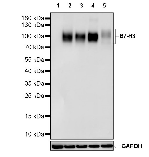
WB result of B7-H3 Recombinant Rabbit mAb
Primary antibody: B7-H3 Recombinant Rabbit mAb at 1/1000 dilution
Lane 1: Raji whole cell lysate 20 µg
Lane 2: unboiled LNCaP whole cell lysate 20 µg
Lane 3: unboiled HEK-293 whole cell lysate 20 µg
Lane 4: U-2 OS whole cell lysate 20 µg
Lane 5: PC-3 whole cell lysate 20 µg
Negative control: Raji whole cell lysate
Secondary antibody: Goat Anti-rabbit IgG, (H+L), HRP conjugated at 1/10000 dilution
Predicted MW: 90 kDa
Observed MW: 90~110 kDa
IHC shows positive staining in paraffin-embedded human tonsil. Anti- B7-H3 antibody was used at 1/100 dilution, followed by a HRP Polymer for Mouse & Rabbit IgG (ready to use). Counterstained with hematoxylin. Heat mediated antigen retrieval with Tris/EDTA buffer pH9.0 was performed before commencing with IHC staining protocol.
IHC shows positive staining in paraffin-embedded human liver. Anti- B7-H3 antibody was used at 1/100 dilution, followed by a HRP Polymer for Mouse & Rabbit IgG (ready to use). Counterstained with hematoxylin. Heat mediated antigen retrieval with Tris/EDTA buffer pH9.0 was performed before commencing with IHC staining protocol.
IHC shows positive staining in paraffin-embedded human prostatic hyperplasia. Anti- B7-H3 antibody was used at 1/100 dilution, followed by a HRP Polymer for Mouse & Rabbit IgG (ready to use). Counterstained with hematoxylin. Heat mediated antigen retrieval with Tris/EDTA buffer pH9.0 was performed before commencing with IHC staining protocol.
IHC shows positive staining in paraffin-embedded human colon cancer. Anti- B7-H3 antibody was used at 1/100 dilution, followed by a HRP Polymer for Mouse & Rabbit IgG (ready to use). Counterstained with hematoxylin. Heat mediated antigen retrieval with Tris/EDTA buffer pH9.0 was performed before commencing with IHC staining protocol.
IHC shows positive staining in paraffin-embedded human prostatic cancer. Anti- B7-H3 antibody was used at 1/100 dilution, followed by a HRP Polymer for Mouse & Rabbit IgG (ready to use). Counterstained with hematoxylin. Heat mediated antigen retrieval with Tris/EDTA buffer pH9.0 was performed before commencing with IHC staining protocol.
Negative control: IHC shows negative staining in paraffin-embedded human skeletal muscle. Anti- B7-H3 antibody was used at 1/100 dilution, followed by a HRP Polymer for Mouse & Rabbit IgG (ready to use). Counterstained with hematoxylin. Heat mediated antigen retrieval with Tris/EDTA buffer pH9.0 was performed before commencing with IHC staining protocol.
 您现在的位置: