
PBS, 40% Glycerol, 0.05% BSA, 0.03% Proclin 300
12 months from date of receipt / reconstitution, -20 °C as supplied
| 应用 | 稀释度 |
|---|---|
| WB | 1:500 |
| IP | 1:50 |
| IHC-P | 1:1000-1:2000 |
| ICC | 1:500 |
| ICFCM | 1:500 |
| ChIP | 1:20~1:50 |
H3K79me1, which stands for Histone H3 monomethylated at Lysine 79, is a specific post-translational modification of histone H3 that involves the addition of a single methyl group to the lysine residue at position 79. It plays a pivotal role in DNA damage response and is involved in various repair mechanisms such as nucleotide excision repair and heterochromatin formation.
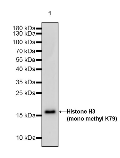
WB result of Histone H3 (mono methyl K79) Recombinant Rabbit mAb
Primary antibody: Histone H3 (mono methyl K79) Recombinant mAb at 1/500 dilution
Lane 1: HeLa whole cell lysate 20 µg
Secondary antibody: Goat Anti-rabbit IgG, (H+L), HRP conjugated at 1/10000 dilution
Predicted MW: 15 kDa
Observed MW: 17 kDa
WB result of Histone H3 (mono methyl K79) Recombinant Rabbit mAb
Primary antibody: Histone H3 (mono methyl K79) Recombinant mAb at 1/500 dilution
Lane 1: NIH/3T3 whole cell lysate 20 µg
Secondary antibody: Goat Anti-rabbit IgG, (H+L), HRP conjugated at 1/10000 dilution
Predicted MW: 15 kDa
Observed MW: 17 kDa
Flow cytometric analysis of 4% PFA fixed 90% methanol permeabilized HeLa (Human cervix adenocarcinoma epithelial cell) labelling Histone H3 (mono methyl K79) antibody at 1/500 dilution (0.1 μg)/ (Red) compared with a Rabbit monoclonal IgG (Black) isotype control and an unlabelled control (cells without incubation with primary antibody and secondary antibody) (Blue). Goat Anti - Rabbit IgG Alexa Fluor® 488 was used as the secondary antibody.
Flow cytometric analysis of 4% PFA fixed 90% methanol permeabilized NIH/3T3 (Mouse embryonic fibroblast) labelling Histone H3 (mono methyl K79) antibody at 1/500 dilution (0.1 μg)/ (Red) compared with a Rabbit monoclonal IgG (Black) isotype control and an unlabelled control (cells without incubation with primary antibody and secondary antibody) (Blue). Goat Anti - Rabbit IgG Alexa Fluor® 488 was used as the secondary antibody.
Histone H3 (mono methyl K79) Rabbit mAb at 1/50 dilution (1 µg) immunoprecipitating Histone H3 (mono methyl K79) in 0.4 mg HeLa whole cell lysate.
Western blot was performed on the immunoprecipitate using Histone H3 (mono methyl K79) Rabbit mAb at 1/1000 dilution.
Secondary antibody (HRP) for IP was used at 1/1000 dilution.
Lane 1: HeLa whole cell lysate 20 µg (Input)
Lane 2: Histone H3 (mono methyl K79) Rabbit mAb IP in HeLa whole cell lysate
Lane 3: Rabbit monoclonal IgG IP in HeLa whole cell lysate
Predicted MW: 15 kDa
Observed MW: 17 kDa
IHC shows positive staining in paraffin-embedded human testis. Anti- Histone H3 (mono methyl K79) antibody was used at 1/1000 dilution, followed by a HRP Polymer for Mouse & Rabbit IgG (ready to use). Counterstained with hematoxylin. Heat mediated antigen retrieval with Tris/EDTA buffer pH9.0 was performed before commencing with IHC staining protocol.
IHC shows positive staining in paraffin-embedded human stomach. Anti- Histone H3 (mono methyl K79) antibody was used at 1/1000 dilution, followed by a HRP Polymer for Mouse & Rabbit IgG (ready to use). Counterstained with hematoxylin. Heat mediated antigen retrieval with Tris/EDTA buffer pH9.0 was performed before commencing with IHC staining protocol.
IHC shows positive staining in paraffin-embedded mouse testis. Anti- Histone H3 (mono methyl K79) antibody was used at 1/2000 dilution, followed by a HRP Polymer for Mouse & Rabbit IgG (ready to use). Counterstained with hematoxylin. Heat mediated antigen retrieval with Tris/EDTA buffer pH9.0 was performed before commencing with IHC staining protocol.
IHC shows positive staining in paraffin-embedded mouse stomach. Anti- Histone H3 (mono methyl K79) antibody was used at 1/2000 dilution, followed by a HRP Polymer for Mouse & Rabbit IgG (ready to use). Counterstained with hematoxylin. Heat mediated antigen retrieval with Tris/EDTA buffer pH9.0 was performed before commencing with IHC staining protocol.
IHC shows positive staining in paraffin-embedded rat testis. Anti- Histone H3 (mono methyl K79) antibody was used at 1/2000 dilution, followed by a HRP Polymer for Mouse & Rabbit IgG (ready to use). Counterstained with hematoxylin. Heat mediated antigen retrieval with Tris/EDTA buffer pH9.0 was performed before commencing with IHC staining protocol.
IHC shows positive staining in paraffin-embedded rat colon. Anti- Histone H3 (mono methyl K79) antibody was used at 1/2000 dilution, followed by a HRP Polymer for Mouse & Rabbit IgG (ready to use). Counterstained with hematoxylin. Heat mediated antigen retrieval with Tris/EDTA buffer pH9.0 was performed before commencing with IHC staining protocol.
ICC shows positive staining in HeLa cells. Anti- Histone H3 (mono methyl K79) antibody was used at 1/500 dilution (Green) and incubated overnight at 4°C. Goat polyclonal Antibody to Rabbit IgG - H&L (Alexa Fluor® 488) was used as secondary antibody at 1/1000 dilution. The cells were fixed with 100% ice-cold methanol and permeabilized with 0.1% PBS-Triton X-100. Nuclei were counterstained with DAPI (Blue). Counterstain with tubulin (Red).
ICC shows positive staining in NIH/3T3 cells. Anti-Histone H3 (mono methyl K79) antibody was used at 1/500 dilution (Green) and incubated overnight at 4°C. Goat polyclonal Antibody to Rabbit IgG - H&L (Alexa Fluor® 488) was used as secondary antibody at 1/1000 dilution. The cells were fixed with 100% ice-cold methanol and permeabilized with 0.1% PBS-Triton X-100. Nuclei were counterstained with DAPI (Blue). Counterstain with tubulin (Red).
Chromatin immunoprecipitation (ChIP) was
performed on HeLa cells cross - linked with 1%
formaldehyde for 10 min, then chromatin was
fragmented by sonication. Parallel reactions used
Histone H3 (mono methyl K79) Recombinant Rabbit mAb
(S-R417) and Rabbit mAb IgG Isotype Control (SDT-R173)
at 1:50 for immunoprecipitation.
Post - immunoprecipitation, both samples were washed,
eluted, and cross - links reversed. Purified DNA was
analyzed by qPCR.
qPCR (%input: immunoprecipitated DNA/input DNA)
showed the enrichment of RPL30, GAPDH, MYOD1, AFM,
SAT-α and SAT-2 in Histone H3 (mono methyl K79)
Recombinant Rabbit mAb (S-R417)-immunoprecipitated
sample.
 您现在的位置: