
12 months from date of receipt / reconstitution, -20 °C as supplied
| 应用 | 稀释度 |
|---|---|
| WB | 1:1000 |
| IHC-P | 1:250 |
| ICC | 1:500 |
| ICFCM | 1:500 |
| IP | 1:50 |
PRMT1 protein, or Protein Arginine Methyltransferase 1, is a key enzyme in cellular biology with diverse functions. PRMT1 catalyzes the symmetric dimethylation of arginine residues in proteins, affecting the structure and function of these proteins. It is involved in multiple biological processes, including cell differentiation, RNA processing, nucleolar formation, and gene transcription and post-transcriptional modification. PRMT1 has been linked to the regulation of miRNA biogenesis by methylating miRNA synthesis-related proteins. PRMT1 is implicated in a wide range of diseases due to its abnormal expression or dysfunction. It has been associated with leukemia, tumors, cardiovascular diseases, and multiple cancer types, including breast cancer, lung cancer, colon cancer, bladder cancer, and blood cancer.
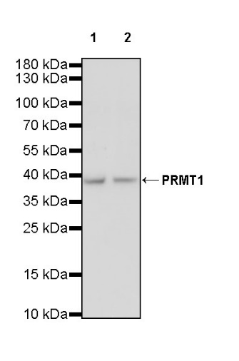
WB result of PRMT1 Recombinant Rabbit mAb
Primary antibody: PRMT1 Recombinant Rabbit mAb at 1/1000 dilution
Lane 1: HepG2 whole cell lysate 20 µg
Lane 2: Jurkat whole cell lysate 20 µg
Secondary antibody: Goat Anti-rabbit IgG, (H+L), HRP conjugated at 1/10000 dilution
Predicted MW: 42 kDa
Observed MW: 38 kDa
WB result of PRMT1 Recombinant Rabbit mAb
Primary antibody: PRMT1 Recombinant Rabbit mAb at 1/1000 dilution
Lane 1: NIH/3T3 whole cell lysate 20 µg
Secondary antibody: Goat Anti-rabbit IgG, (H+L), HRP conjugated at 1/10000 dilution
Predicted MW: 42 kDa
Observed MW: 38 kDa
WB result of PRMT1 Recombinant Rabbit mAb
Primary antibody: PRMT1 Recombinant Rabbit mAb at 1/1000 dilution
Lane 1: rat brain lysate 20 µg
Secondary antibody: Goat Anti-rabbit IgG, (H+L), HRP conjugated at 1/10000 dilution
Predicted MW: 42 kDa
Observed MW: 38 kDa
This blot was developed with high sensitivity substrate
Flow cytometric analysis of 4% PFA fixed 90% methanol permeabilized HepG2 (Human hepatocellular carcinoma epithelial cell) labelling PRMT1 antibody at 1/500 dilution (0.1 μg)/ (Red) compared with a Rabbit monoclonal IgG (Black) isotype control and an unlabelled control (cells without incubation with primary antibody and secondary antibody) (Blue). Goat Anti - Rabbit IgG Alexa Fluor® 488 was used as the secondary antibody.
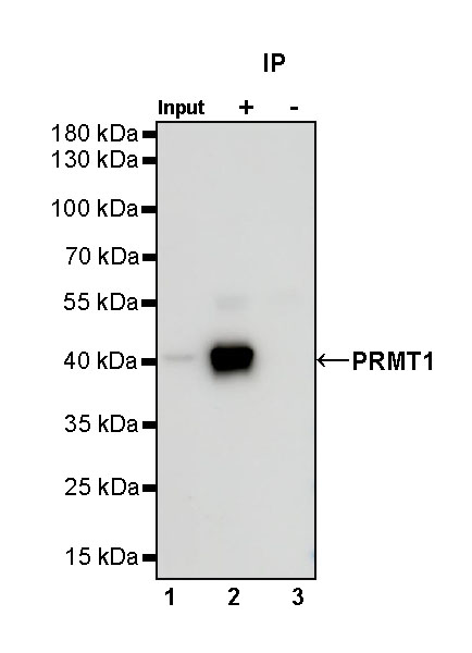
PRMT1 Rabbit mAb at 1/50 dilution (1 µg) immunoprecipitating PRMT1 in 0.4 mg HepG2 whole cell lysate.
Western blot was performed on the immunoprecipitate using PRMT1 Rabbit mAb at 1/1000 dilution.
Secondary antibody (HRP) for IP was used at 1/1000 dilution.
Lane 1: HepG2 whole cell lysate 20 µg (Input)
Lane 2: PRMT1 Rabbit mAb IP in HepG2 whole cell lysate
Lane 3: Rabbit monoclonal IgG IP in HepG2 whole cell lysate
Predicted MW: 42 kDa
Observed MW: 38 kDa
IHC shows positive staining in paraffin-embedded human testis. Anti-PRMT1 antibody was used at 1/250 dilution, followed by a HRP Polymer for Mouse & Rabbit IgG (ready to use). Counterstained with hematoxylin. Heat mediated antigen retrieval with Tris/EDTA buffer pH9.0 was performed before commencing with IHC staining protocol.
Negative control: IHC shows negative staining in paraffin-embedded human cardiac muscle. Anti-PRMT1 antibody was used at 1/250 dilution, followed by a HRP Polymer for Mouse & Rabbit IgG (ready to use). Counterstained with hematoxylin. Heat mediated antigen retrieval with Tris/EDTA buffer pH9.0 was performed before commencing with IHC staining protocol.
IHC shows positive staining in paraffin-embedded human colon cancer. Anti-PRMT1 antibody was used at 1/250 dilution, followed by a HRP Polymer for Mouse & Rabbit IgG (ready to use). Counterstained with hematoxylin. Heat mediated antigen retrieval with Tris/EDTA buffer pH9.0 was performed before commencing with IHC staining protocol.
IHC shows positive staining in paraffin-embedded human breast cancer. Anti-PRMT1 antibody was used at 1/250 dilution, followed by a HRP Polymer for Mouse & Rabbit IgG (ready to use). Counterstained with hematoxylin. Heat mediated antigen retrieval with Tris/EDTA buffer pH9.0 was performed before commencing with IHC staining protocol.
IHC shows positive staining in paraffin-embedded human colon. Anti-PRMT1 antibody was used at 1/250 dilution, followed by a HRP Polymer for Mouse & Rabbit IgG (ready to use). Counterstained with hematoxylin. Heat mediated antigen retrieval with Tris/EDTA buffer pH9.0 was performed before commencing with IHC staining protocol.
ICC shows positive staining in HepG2 cells. Anti-PRMT1 antibody was used at 1/500 dilution (Green) and incubated overnight at 4°C. Goat polyclonal Antibody to Rabbit IgG - H&L (Alexa Fluor® 488) was used as secondary antibody at 1/1000 dilution. The cells were fixed with 4% PFA and permeabilized with 0.1% PBS-Triton X-100. Nuclei were counterstained with DAPI (Blue). Counterstain with tubulin (Red).
 您现在的位置: