
PBS, 40% Glycerol, 0.05% BSA, 0.03% Proclin 300
12 months from date of receipt / reconstitution, -20 °C as supplied
| 应用 | 稀释度 |
|---|---|
| WB | 1:5000 |
| IP | 1:50 |
| ICC | 1:500 |
The E tag is an epitope tag with a 13-residue sequence of GAPVPYPDPLEPR. The E tag can help with detection of proteins in western blot, IP, or microscopic techniques.
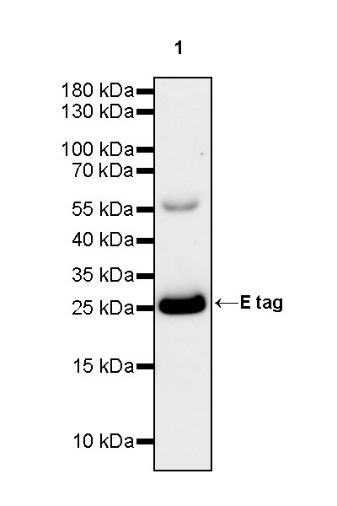
WB result of E tag Recombinant Rabbit mAb
Primary antibody: E tag Recombinant Rabbit mAb at 1/5000 dilution
Lane 1: E tag protein 10 ng
Secondary antibody: Goat Anti-rabbit IgG, (H+L), HRP conjugated at 1/10000 dilution
Predicted MW: 26 kDa
Observed MW: 26 kDa
E tag Rabbit mAb at 1/50 dilution (1 µg) immunoprecipitating E tag in 0.4 mg 293T transfected with Histone H3-mCherry-E-tag whole cell lysate.
Western blot was performed on the immunoprecipitate using E tag Rabbit mAb at 1/1000 dilution.
Secondary antibody (HRP) for IP was used at 1/1000 dilution.
Lane 1: 293T transfected with Histone H3-mCherry-E-tag whole cell lysate 5 µg (Input)
Lane 2: E tag Rabbit mAb IP in 293T transfected with Histone H3-mCherry-E-tag whole cell lysate
Lane 3: Rabbit monoclonal IgG IP in 293T transfected with Histone H3-mCherry-E-tag whole cell lysate
Predicted MW: 26 kDa
Observed MW: 26,50 kDa
ICC shows positive staining in Histone H3-mCherry-E-tag transfected 293T cells. Anti-E tag antibody was used at 1/500 dilution (Green) and incubated overnight at 4°C. Goat polyclonal Antibody to Rabbit IgG - H&L (Alexa Fluor® 488) was used as secondary antibody at 1/1000 dilution. The cells were fixed with 4% PFA and permeabilized with 0.1% PBS-Triton X-100. Nuclei were counterstained with DAPI (Blue).
Negative control:ICC shows negative staining in vector-transfected 293T cells. Anti-E tag antibody was used at 1/500 dilution and incubated overnight at 4°C. Goat polyclonal Antibody to Rabbit IgG - H&L (Alexa Fluor® 488) was used as secondary antibody at 1/1000 dilution. The cells were fixed with 4% PFA and permeabilized with 0.1% PBS-Triton X-100. Nuclei were counterstained with DAPI (Blue).
 您现在的位置: