
12 months from date of receipt / reconstitution, -20 °C as supplied
| 应用 | 稀释度 |
|---|---|
| WB | 1:1000 |
| IP | 1:50 |
| IHC-P | 1:250 |
| ICC | 1:500 |
| ICFCM | 1:50 |
JunB is a member of the Jun family of proteins, which belongs to the larger group of transcription factors known as the AP-1 (Activator Protein 1) complex. AP-1 complexes are dimers composed of Jun (c-Jun, JunB, JunD) and Fos (c-Fos, FosB, Fra1, Fra2) proteins. These dimers regulate gene expression by binding to specific DNA sequences called AP-1 response elements in the promoter regions of target genes. JunB plays a crucial role in cell proliferation, differentiation, apoptosis, and other cellular processes. It can function as both a transcriptional activator and repressor, depending on the cellular context and the interacting proteins. JunB is also involved in regulating the immune response and inflammation. In cancer research, JunB has been found to have both tumor-suppressing and tumor-promoting roles. It can act as a tumor suppressor by inhibiting cell proliferation and promoting apoptosis, but it can also promote tumor growth and invasion by activating genes that are involved in cell migration and invasion. The expression and activity of JunB are tightly regulated by various signaling pathways, including the MAPK (Mitogen-Activated Protein Kinase) pathway. Aberrant JunB expression or activity has been associated with a variety of diseases, including cancer, autoimmune diseases, and inflammatory disorders.
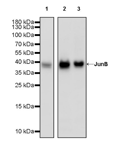
WB result of JunB Recombinant Rabbit mAb
Primary antibody: JunB Recombinant Rabbit mAb at 1/1000 dilution
Lane 1: HeLa whole cell lysate 20 µg
Lane 2: A431 whole cell lysate 20 µg
Lane 3: PC-3 whole cell lysate 20 µg
Secondary antibody: Goat Anti-rabbit IgG, (H+L), HRP conjugated at 1/10000 dilution
Predicted MW: 36 kDa
Observed MW: 38 kDa
WB result of JunB Recombinant Rabbit mAb
Primary antibody: JunB Recombinant Rabbit mAb at 1/1000 dilution
Lane 1: NIH/3T3 whole cell lysate 20 µg
Secondary antibody: Goat Anti-rabbit IgG, (H+L), HRP conjugated at 1/10000 dilution
Predicted MW: 36 kDa
Observed MW: 38 kDa
WB result of JunB Recombinant Rabbit mAb
Primary antibody: JunB Recombinant Rabbit mAb at 1/1000 dilution
Lane 1: C6 whole cell lysate 20 µg
Secondary antibody: Goat Anti-rabbit IgG, (H+L), HRP conjugated at 1/10000 dilution
Predicted MW: 36 kDa
Observed MW: 38 kDa
Flow cytometric analysis of 4% PFA fixed 90% methanol permeabilized HeLa (Human cervix adenocarcinoma epithelial cell) labelling JunB antibody at 1/50 dilution (1 μg)/ (Red) compared with a Rabbit monoclonal IgG (Black) isotype control and an unlabelled control (cells without incubation with primary antibody and secondary antibody) (Blue). Goat Anti - Rabbit IgG Alexa Fluor® 488 was used as the secondary antibody.
JunB Rabbit mAb at 1/50 dilution (1 µg) immunoprecipitating JunB in 0.4 mg PC-3 whole cell lysate.
Western blot was performed on the immunoprecipitate using JunB Rabbit mAb at 1/1000 dilution.
Secondary antibody (HRP) for IP was used at 1/1000 dilution.
Lane 1: PC-3 whole cell lysate 20 µg (Input)
Lane 2: JunB Rabbit mAb IP in PC-3 whole cell lysate
Lane 3: Rabbit monoclonal IgG IP in PC-3 whole cell lysate
Predicted MW: 36 kDa
Observed MW: 38 kDa
IHC shows positive staining in paraffin-embedded human stomach. Anti-JunB antibody was used at 1/250 dilution, followed by a HRP Polymer for Mouse & Rabbit IgG (ready to use). Counterstained with hematoxylin. Heat mediated antigen retrieval with Tris/EDTA buffer pH9.0 was performed before commencing with IHC staining protocol.
IHC shows positive staining in paraffin-embedded human tonsil. Anti-JunB antibody was used at 1/250 dilution, followed by a HRP Polymer for Mouse & Rabbit IgG (ready to use). Counterstained with hematoxylin. Heat mediated antigen retrieval with Tris/EDTA buffer pH9.0 was performed before commencing with IHC staining protocol.
IHC shows positive staining in paraffin-embedded mouse stomach. Anti-JunB antibody was used at 1/250 dilution, followed by a HRP Polymer for Mouse & Rabbit IgG (ready to use). Counterstained with hematoxylin. Heat mediated antigen retrieval with Tris/EDTA buffer pH9.0 was performed before commencing with IHC staining protocol.
IHC shows positive staining in paraffin-embedded rat spleen. Anti-JunB antibody was used at 1/250 dilution, followed by a HRP Polymer for Mouse & Rabbit IgG (ready to use). Counterstained with hematoxylin. Heat mediated antigen retrieval with Tris/EDTA buffer pH9.0 was performed before commencing with IHC staining protocol.
ICC shows positive staining in HeLa cells. Anti-JunB antibody was used at 1/500 dilution (Green) and incubated overnight at 4°C. Goat polyclonal Antibody to Rabbit IgG - H&L (Alexa Fluor® 488) was used as secondary antibody at 1/1000 dilution. The cells were fixed with 4% PFA and permeabilized with 0.1% PBS-Triton X-100. Nuclei were counterstained with DAPI (Blue). Counterstain with tubulin (Red).
 您现在的位置: