
12 months from date of receipt / reconstitution, -20 °C as supplied
| 应用 | 稀释度 |
|---|---|
| WB | 1:1000 |
| IP | 1:50 |
| IHC-P | 1:250 |
| ICC | 1:50 |
| ICFCM | 1:500 |
KIF3A is a subunit of the heterotrimeric kinesin-2 complex, which belongs to the N-kinesin family of microtubule plus-end-directed motor proteins. Along with KIF3B and the cargo-binding protein KAP3, it forms the heterotrimeric kinesin-2 complex that is involved in regulating intracellular transport and mediating microtubule-based motility. The specific functions of KIF3A include roles in regulating bone formation, nephron number, cell survival, and gene expression control. It is also implicated in sperm formation, prostate cancer suppression, and glioblastoma, among others. KIF3A is associated with diseases such as Usher syndrome type I and neuronal ceroid lipofuscinosis.
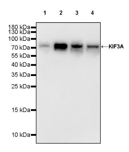
WB result of KIF3A Recombinant Rabbit mAb
Primary antibody: KIF3A Recombinant Rabbit mAb at 1/1000 dilution
Lane 1: HeLa whole cell lysate 20 µg
Lane 2: SH-SY5Y whole cell lysate 20 µg
Lane 3: 293T whole cell lysate 20 µg
Lane 4: MDA-MB-231 whole cell lysate 20 µg
Secondary antibody: Goat Anti-rabbit IgG, (H+L), HRP conjugated at 1/10000 dilution
Predicted MW: 80 kDa
Observed MW: 80 kDa
This blot was developed with high sensitivity substrate
WB result of KIF3A Recombinant Rabbit mAb
Primary antibody: KIF3A Recombinant Rabbit mAb at 1/1000 dilution
Lane 1: mouse brain lysate 20 µg
Secondary antibody: Goat Anti-rabbit IgG, (H+L), HRP conjugated at 1/10000 dilution
Predicted MW: 80 kDa
Observed MW: 80 kDa
This blot was developed with high sensitivity substrate
WB result of KIF3A Recombinant Rabbit mAb
Primary antibody: KIF3A Recombinant Rabbit mAb at 1/1000 dilution
Lane 1: rat brain lysate 20 µg
Secondary antibody: Goat Anti-rabbit IgG, (H+L), HRP conjugated at 1/10000 dilution
Predicted MW: 80 kDa
Observed MW: 80 kDa
This blot was developed with high sensitivity substrate
Flow cytometric analysis of 4% PFA fixed 90% methanol permeabilized HeLa (Human cervix adenocarcinoma epithelial cell) labelling KIF3A antibody at 1/500 dilution (0.1 μg)/ (Red) compared with a Rabbit monoclonal IgG (Black) isotype control and an unlabelled control (cells without incubation with primary antibody and secondary antibody) (Blue). Goat Anti - Rabbit IgG Alexa Fluor® 488 was used as the secondary antibody.
KIF3A Rabbit mAb at 1/50 dilution (1 µg) immunoprecipitating KIF3A in 0.4 mg SH-SY5Y whole cell lysate.
Western blot was performed on the immunoprecipitate using KIF3A Rabbit mAb at 1/1000 dilution.
Secondary antibody (HRP) for IP was used at 1/1000 dilution.
Lane 1: SH-SY5Y whole cell lysate 20 µg (Input)
Lane 2: KIF3A Rabbit mAb IP in SH-SY5Y whole cell lysate
Lane 3: Rabbit monoclonal IgG IP in SH-SY5Y whole cell lysate
Predicted MW: 80 kDa
Observed MW: 80 kDa
IHC shows positive staining in paraffin-embedded human testis. Anti-KIF3A antibody was used at 1/250 dilution, followed by a HRP Polymer for Mouse & Rabbit IgG (ready to use). Counterstained with hematoxylin. Heat mediated antigen retrieval with Tris/EDTA buffer pH9.0 was performed before commencing with IHC staining protocol.
IHC shows positive staining in paraffin-embedded human cerebellum. Anti-KIF3A antibody was used at 1/250 dilution, followed by a HRP Polymer for Mouse & Rabbit IgG (ready to use). Counterstained with hematoxylin. Heat mediated antigen retrieval with Tris/EDTA buffer pH9.0 was performed before commencing with IHC staining protocol.
IHC shows positive staining in paraffin-embedded mouse cerebral cortex. Anti-KIF3A antibody was used at 1/250 dilution, followed by a HRP Polymer for Mouse & Rabbit IgG (ready to use). Counterstained with hematoxylin. Heat mediated antigen retrieval with Tris/EDTA buffer pH9.0 was performed before commencing with IHC staining protocol.
IHC shows positive staining in paraffin-embedded mouse cerebellum. Anti-KIF3A antibody was used at 1/250 dilution, followed by a HRP Polymer for Mouse & Rabbit IgG (ready to use). Counterstained with hematoxylin. Heat mediated antigen retrieval with Tris/EDTA buffer pH9.0 was performed before commencing with IHC staining protocol.
IHC shows positive staining in paraffin-embedded rat cerebral cortex. Anti-KIF3A antibody was used at 1/250 dilution, followed by a HRP Polymer for Mouse & Rabbit IgG (ready to use). Counterstained with hematoxylin. Heat mediated antigen retrieval with Tris/EDTA buffer pH9.0 was performed before commencing with IHC staining protocol.
IHC shows positive staining in paraffin-embedded rat cerebellum. Anti-KIF3A antibody was used at 1/250 dilution, followed by a HRP Polymer for Mouse & Rabbit IgG (ready to use). Counterstained with hematoxylin. Heat mediated antigen retrieval with Tris/EDTA buffer pH9.0 was performed before commencing with IHC staining protocol.
ICC shows positive staining in SH-SY5Y cells. Anti-KIF3A antibody was used at 1/50 dilution (Green) and incubated overnight at 4°C. Goat polyclonal Antibody to Rabbit IgG - H&L (Alexa Fluor® 488) was used as secondary antibody at 1/1000 dilution. The cells were fixed with 4% PFA and permeabilized with 0.1% PBS-Triton X-100. Nuclei were counterstained with DAPI (Blue). Counterstain with tubulin (Red).
 您现在的位置: