
12 months from date of receipt / reconstitution, -20 °C as supplied
| 应用 | 稀释度 |
|---|---|
| WB | 1:1000 |
| IP | 1:50 |
| IHC-P | 1:100 |
| ICC | 1:500 |
| ICFCM | 1:500 |
TOP1 is a DNA topoisomerase, an enzyme that controls and alters the topologic states of DNA during transcription. This enzyme catalyzes the transient breaking and rejoining of a single strand of DNA which lets the broken strand rotate around the intact strand, thus altering the topology of DNA. The eukaryotic topoisomerases I were found to nick the DNA with a preference for a sequence of nucleotides that extends from positions -4 to -1 from the nick. The preferred nucleotides in the strand to be cut are 5'-(A/T)(G/C)(A/T)T-3' with the enzyme covalently attached to the -1 T residue, though sometimes a C residue is found at the -1 position. TOP1 has been known as a target for the treatment of human cancers. Camptothecin analogues irinotecan and topotecan, which inhibit TOP1, are among the most effective FDA-approved anticancer chemotherapeutic agents used in clinical practice. Higher expression of TOP1 in KRAS mutant non-small cell lung cancer and correlation to survival suggests that TOP1 inhibitors might have increased benefit when administered to treat patients with a KRAS mutant tumor.
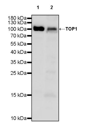
WB result of TOP1 Recombinant Rabbit mAb
Primary antibody: TOP1 Recombinant Rabbit mAb at 1/1000 dilution
Lane 1: MCF7 whole cell lysate 20 µg
Lane 2: HepG2 whole cell lysate 20 µg
Secondary antibody: Goat Anti-rabbit IgG, (H+L), HRP conjugated at 1/10000 dilution
Predicted MW: 91 kDa
Observed MW: 100 kDa
WB result of TOP1 Recombinant Rabbit mAb
Primary antibody: TOP1 Recombinant Rabbit mAb at 1/1000 dilution
Lane 1: Neuro-2a whole cell lysate 20 µg
Secondary antibody: Goat Anti-rabbit IgG, (H+L), HRP conjugated at 1/10000 dilution
Predicted MW: 91 kDa
Observed MW: 100 kDa
WB result of TOP1 Recombinant Rabbit mAb
Primary antibody: TOP1 Recombinant Rabbit mAb at 1/1000 dilution
Lane 1: PC-12 whole cell lysate 20 µg
Secondary antibody: Goat Anti-rabbit IgG, (H+L), HRP conjugated at 1/10000 dilution
Predicted MW: 91 kDa
Observed MW: 100 kDa
Flow cytometric analysis of 4% PFA fixed 90% methanol permeabilized MCF-7 (Human breast adenocarcinoma epithelial cell) labelling TOP1 antibody at 1/500 dilution (0.1 μg)/ (Red) compared with a Rabbit monoclonal IgG (Black) isotype control and an unlabelled control (cells without incubation with primary antibody and secondary antibody) (Blue). Goat Anti - Rabbit IgG Alexa Fluor® 488 was used as the secondary antibody.
TOP1 Rabbit mAb at 1/50 dilution (1 µg) immunoprecipitating TOP1 in 0.4 mg MCF7 whole cell lysate.
Western blot was performed on the immunoprecipitate using TOP1 Rabbit mAb at 1/1000 dilution.
Secondary antibody (HRP) for IP was used at 1/1000 dilution.
Lane 1: MCF7 whole cell lysate 20 µg (Input)
Lane 2: TOP1 Rabbit mAb IP in MCF7 whole cell lysate
Lane 3: Rabbit monoclonal IgG IP in MCF7 whole cell lysate
Predicted MW: 91 kDa
Observed MW: 100 kDa
IHC shows positive staining in paraffin-embedded human stomach. Anti-TOP1 antibody was used at 1/100 dilution, followed by a HRP Polymer for Mouse & Rabbit IgG (ready to use). Counterstained with hematoxylin. Heat mediated antigen retrieval with Tris/EDTA buffer pH9.0 was performed before commencing with IHC staining protocol.
IHC shows positive staining in paraffin-embedded human testis. Anti-TOP1 antibody was used at 1/100 dilution, followed by a HRP Polymer for Mouse & Rabbit IgG (ready to use). Counterstained with hematoxylin. Heat mediated antigen retrieval with Tris/EDTA buffer pH9.0 was performed before commencing with IHC staining protocol.
IHC shows positive staining in paraffin-embedded human cervical squamous cell carcinoma. Anti-TOP1 antibody was used at 1/100 dilution, followed by a HRP Polymer for Mouse & Rabbit IgG (ready to use). Counterstained with hematoxylin. Heat mediated antigen retrieval with Tris/EDTA buffer pH9.0 was performed before commencing with IHC staining protocol.
IHC shows positive staining in paraffin-embedded human endometrial carcinoma. Anti-TOP1 antibody was used at 1/100 dilution, followed by a HRP Polymer for Mouse & Rabbit IgG (ready to use). Counterstained with hematoxylin. Heat mediated antigen retrieval with Tris/EDTA buffer pH9.0 was performed before commencing with IHC staining protocol.
IHC shows positive staining in paraffin-embedded rat colon. Anti-TOP1 antibody was used at 1/100 dilution, followed by a HRP Polymer for Mouse & Rabbit IgG (ready to use). Counterstained with hematoxylin. Heat mediated antigen retrieval with Tris/EDTA buffer pH9.0 was performed before commencing with IHC staining protocol.
IHC shows positive staining in paraffin-embedded rat kidney. Anti-TOP1 antibody was used at 1/100 dilution, followed by a HRP Polymer for Mouse & Rabbit IgG (ready to use). Counterstained with hematoxylin. Heat mediated antigen retrieval with Tris/EDTA buffer pH9.0 was performed before commencing with IHC staining protocol.
ICC shows positive staining in MCF-7 cells. Anti-TOP1 antibody was used at 1/500 dilution (Green) and incubated overnight at 4°C. Goat polyclonal Antibody to Rabbit IgG - H&L (Alexa Fluor® 488) was used as secondary antibody at 1/1000 dilution. The cells were fixed with 4% PFA and permeabilized with 0.1% PBS-Triton X-100. Nuclei were counterstained with DAPI (Blue). Counterstain with tubulin (Red).
 您现在的位置: