
PBS, 0.1% BSA, 0.01% Proclin 300
12 months from date of receipt / reconstitution, 2 to 8 °C as supplied
Isotype control antibodies, to estimate the nonspecific binding of target. Use at concentrations comparable to those of the specific antibody of interest.
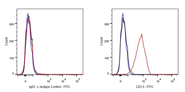
Flow cytometric analysis of THP-1 (Human monocytic leukemia monocyte) labeling SDT FITC Mouse IgG1, κ Isotype Control Antibody at 1 μg/test (Left) / (Red line histogram) compared with SDT CD13 antibody at 1 μg/test (S0B0441) (Right) / (Red line histogram), Mouse monoclonal IgG Isotype Control (Goat Anti - Mouse IgG FITC was used as the secondary antibody) (Left) / (Black line histogram) compared with SDT FITC Mouse IgG1, κ Isotype Control Antibody (Right) / (Black line histogram) and an unlabelled control (cells without incubation with primary antibody and secondary antibody) (Blue line histogram). Flow cytometry and data analysis were performed using BD FACSymphony™ A1 and FlowJo™ software.
Flow cytometric analysis of 4% paraformaldehyde fixed 90% methanol permeabilized HeLa (Human cervix adenocarcinoma epithelial cell) labelling SDT FITC Mouse IgG1, κ Isotype Control Antibody at 1 μg/test (Left) / (Red line histogram) compared with α-tubulin antibody (Right) / (Red line histogram), Mouse monoclonal IgG Isotype Control (Goat Anti - Mouse IgG FITC was used as the secondary antibody) (Left) / (Black line histogram) compared with SDT FITC Mouse IgG1, κ Isotype Control Antibody (Right) / (Black line histogram) and an unlabelled control (cells without incubation with primary antibody and secondary antibody) (Blue line histogram). Flow cytometry and data analysis were performed using BD FACSymphony™ A1 and FlowJo™ software.
 您现在的位置: