
12 months from date of receipt / reconstitution, -20 °C as supplied
| 应用 | 稀释度 |
|---|---|
| IHC-P | 1:1000 |
| ICC | 1:50 |
| ICFCM | 1:25 |
| IF | 1:500 |
COX-2 protein, also known as Cyclooxygenase-2 or PGHS-2, is a member of the cyclooxygenase superfamily. This recombinant protein plays a specific enzymatic role in the biological system, converting arachidonic acid (AA) into prostaglandin G2 and other peroxide substances, participating in the biosynthesis of prostaglandins. Under normal physiological conditions, most tissue cells do not express COX-2 protein. However, in pathological states such as inflammation and tumorigenesis, COX-2 protein expression is upregulated in response to proinflammatory mediators, including inflammatory stimuli, damage, mitogens, and carcinogens. This process involves the release of arachidonic acid from cell membrane phospholipids through the phospholipase A2 pathway, followed by the synthesis of inflammatory mediators such as prostaglandin E2 (PGE2) catalyzed by COX-2, participating in various physiological and pathological processes in the body. Moreover, the enzymatic activity of COX-2 protein is not limited to arachidonic acid. It can also act on dihomo-γ-linolenic acid (DGLA) and eicosapentaenoic acid (EPA), generating PGH1 and PGH3 as precursors of different series of prostaglandins.
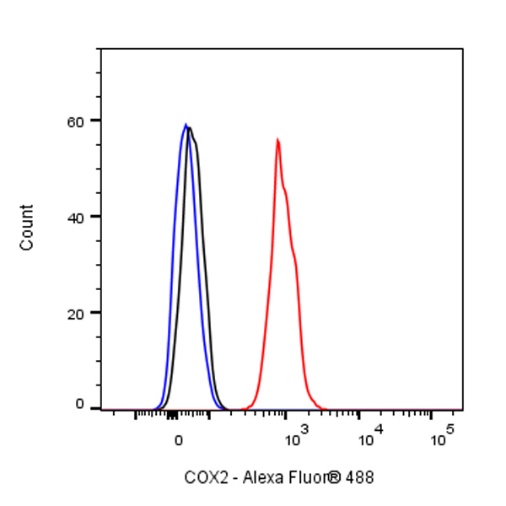
Flow cytometric analysis of 4% PFA fixed 90% methanol permeabilized U87-MG (Human glioblastoma-astrocytoma epithelial cell) labelling COX2 antibody at 1/25 dilution (1 μg)/ (Red) compared with a Rabbit monoclonal IgG (Black) isotype control and an unlabelled control (cells without incubation with primary antibody and secondary antibody) (Blue). Goat Anti - Rabbit IgG Alexa Fluor® 488 was used as the secondary antibody.
IHC shows positive staining in paraffin-embedded human colon. Anti-COX2 antibody was used at 1/1000 dilution, followed by a HRP Polymer for Mouse & Rabbit IgG (ready to use). Counterstained with hematoxylin. Heat mediated antigen retrieval with Tris/EDTA buffer pH9.0 was performed before commencing with IHC staining protocol.
IHC shows positive staining in paraffin-embedded human kidney. Anti-COX2 antibody was used at 1/1000 dilution, followed by a HRP Polymer for Mouse & Rabbit IgG (ready to use). Counterstained with hematoxylin. Heat mediated antigen retrieval with Tris/EDTA buffer pH9.0 was performed before commencing with IHC staining protocol.
IHC shows positive staining in paraffin-embedded human liver. Anti-COX2 antibody was used at 1/1000 dilution, followed by a HRP Polymer for Mouse & Rabbit IgG (ready to use). Counterstained with hematoxylin. Heat mediated antigen retrieval with Tris/EDTA buffer pH9.0 was performed before commencing with IHC staining protocol.
IHC shows positive staining in paraffin-embedded human colon cancer. Anti-COX2 antibody was used at 1/1000 dilution, followed by a HRP Polymer for Mouse & Rabbit IgG (ready to use). Counterstained with hematoxylin. Heat mediated antigen retrieval with Tris/EDTA buffer pH9.0 was performed before commencing with IHC staining protocol.
IHC shows positive staining in paraffin-embedded human renal clear cell carcinoma. Anti-COX2 antibody was used at 1/1000 dilution, followed by a HRP Polymer for Mouse & Rabbit IgG (ready to use). Counterstained with hematoxylin. Heat mediated antigen retrieval with Tris/EDTA buffer pH9.0 was performed before commencing with IHC staining protocol.
ICC shows positive staining in U87-MG cells. Anti-COX2 antibody was used at 1/50 dilution (Green) and incubated overnight at 4°C. Goat polyclonal Antibody to Rabbit IgG - H&L (Alexa Fluor® 488) was used as secondary antibody at 1/1000 dilution. The cells were fixed with 100% ice-cold methanol and permeabilized with 0.1% PBS-Triton X-100. Nuclei were counterstained with DAPI (Blue). Counterstain with tubulin (Red).
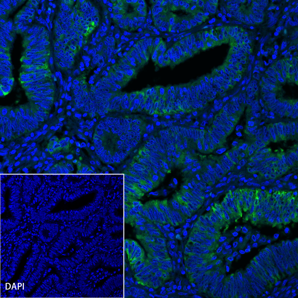
IF shows positive staining in paraffin-embedded human colon cancer. Anti-COX2 antibody was used at 1/500 dilution (Green) and incubated overnight at 4°C. Goat polyclonal Antibody to Rabbit IgG - H&L (Alexa Fluor® 488) (S0B4004) was used as secondary antibody at 1/500 dilution. Counterstained with DAPI (Blue). Heat mediated antigen retrieval with EDTA buffer pH9.0 was performed before commencing with IF staining protocol.
 您现在的位置: