
12 months from date of receipt / reconstitution, -20 °C as supplied
| 应用 | 稀释度 |
|---|---|
| WB | 1:1000 |
| IP | 1:50 |
| IHC-P | 1:250 |
BAD protein, or Bcl-2-associated death promoter, is a member of the Bcl-2 protein family that is involved in the regulation of apoptosis (cell death). It functions as a pro-apoptotic protein, promoting cell death when activated. BAD protein interacts with other Bcl-2 family members, such as Bcl-2 and Bcl-xL, to regulate the release of cytochrome c from the mitochondria, which is a crucial step in the apoptotic cascade. The activity of BAD protein is regulated by phosphorylation. When BAD is phosphorylated, it dissociates from Bcl-2 and Bcl-xL, losing its pro-apoptotic function. Conversely, when BAD is dephosphorylated, it binds to Bcl-2 and Bcl-xL, inhibiting their anti-apoptotic activity and promoting apoptosis. The phosphorylation of BAD protein is mediated by several kinases, including AKT (also known as protein kinase B). AKT phosphorylates BAD at specific sites, preventing its interaction with Bcl-2 and Bcl-xL and thus inhibiting apoptosis. This mechanism is important in promoting cell survival and proliferation in response to growth factors and other stimuli.
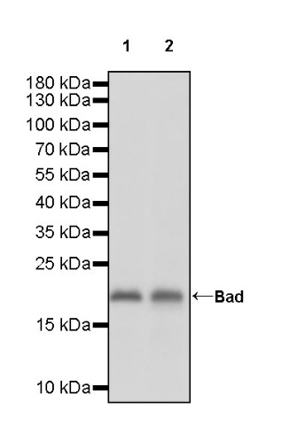
WB result of Bad Rabbit mAb
Primary antibody: Bad Rabbit mAb at 1/1000 dilution
Lane 1: HeLa whole cell lysate 20 µg
Lane 2: MCF7 whole cell lysate 20 µg
Secondary antibody: Goat Anti-rabbit IgG, (H+L), HRP conjugated at 1/10000 dilution
Predicted MW: 18 kDa
Observed MW: 20 kDa
WB result of Bad Rabbit mAb
Primary antibody: Bad Rabbit mAb at 1/1000 dilution
Lane 1: C2C12 whole cell lysate 20 µg
Secondary antibody: Goat Anti-rabbit IgG, (H+L), HRP conjugated at 1/10000 dilution
Predicted MW: 18 kDa
Observed MW: 20 kDa
(This blot was developed with high sensitivity substrate)
WB result of Bad Rabbit mAb
Primary antibody: Bad Rabbit mAb at 1/1000 dilution
Lane 1: C6 whole cell lysate 20 µg
Secondary antibody: Goat Anti-rabbit IgG, (H+L), HRP conjugated at 1/10000 dilution
Predicted MW: 18 kDa
Observed MW: 20 kDa
Bad Rabbit mAb at 1/50 dilution (1 µg) immunoprecipitating Bad in 0.4 mg HeLa whole cell lysate.
Western blot was performed on the immunoprecipitate using Bad Rabbit mAb at 1/1000 dilution.
Secondary antibody (HRP) for IP was used at 1/1000 dilution.
Lane 1: HeLa whole cell lysate 20 µg (Input)
Lane 2: Bad Rabbit mAb IP in HeLa whole cell lysate
Lane 3: Rabbit monoclonal IgG IP in HeLa whole cell lysate
Predicted MW: 18 kDa
Observed MW: 20 kDa
IHC shows positive staining in paraffin-embedded human colon. Anti-Bad antibody was used at 1/250 dilution, followed by a HRP Polymer for Mouse & Rabbit IgG (ready to use). Counterstained with hematoxylin. Heat mediated antigen retrieval with Tris/EDTA buffer pH9.0 was performed before commencing with IHC staining protocol.
IHC shows positive staining in paraffin-embedded human kidney. Anti-Bad antibody was used at 1/250 dilution, followed by a HRP Polymer for Mouse & Rabbit IgG (ready to use). Counterstained with hematoxylin. Heat mediated antigen retrieval with Tris/EDTA buffer pH9.0 was performed before commencing with IHC staining protocol.
Negative control: IHC shows negative staining in paraffin-embedded human skeletal muscle. Anti-Bad antibody was used at 1/250 dilution, followed by a HRP Polymer for Mouse & Rabbit IgG (ready to use). Counterstained with hematoxylin. Heat mediated antigen retrieval with Tris/EDTA buffer pH9.0 was performed before commencing with IHC staining protocol.
IHC shows positive staining in paraffin-embedded human colon cancer. Anti-Bad antibody was used at 1/250 dilution, followed by a HRP Polymer for Mouse & Rabbit IgG (ready to use). Counterstained with hematoxylin. Heat mediated antigen retrieval with Tris/EDTA buffer pH9.0 was performed before commencing with IHC staining protocol.
IHC shows positive staining in paraffin-embedded mouse kidney. Anti-Bad antibody was used at 1/250 dilution, followed by a HRP Polymer for Mouse & Rabbit IgG (ready to use). Counterstained with hematoxylin. Heat mediated antigen retrieval with Tris/EDTA buffer pH9.0 was performed before commencing with IHC staining protocol.
IHC shows positive staining in paraffin-embedded rat kidney. Anti-Bad antibody was used at 1/250 dilution, followed by a HRP Polymer for Mouse & Rabbit IgG (ready to use). Counterstained with hematoxylin. Heat mediated antigen retrieval with Tris/EDTA buffer pH9.0 was performed before commencing with IHC staining protocol.
 您现在的位置: