
PBS, 40% Glycerol, 0.05% BSA, 0.03% Proclin 300
12 months from date of receipt / reconstitution, -20 °C as supplied
| 应用 | 稀释度 |
|---|---|
| WB | 1:1000 |
| ICC | 1:500 |
| FCM | 1:2000 |
CD11c is a type I transmembrane glycoprotein of approximately 150 kDa, also known as integrin alpha X (Itgax). It is part of the complement receptor 4 (CR4) complex and, along with CR3 (also known as Mac-1 or CD11b/CD18), belongs to the β2-integrin family of adhesion molecules. CD11c is primarily expressed on dendritic cells (DCs), NK cells, a subset of intestinal intraepithelial lymphocytes (IELs), and some activated T cells. It is involved in cell adhesion, migration, and phagocytic functions, mediating the phagocytic process of DCs through its association with β2-integrin. Under conditions of early development and in certain neurodegenerative diseases such as Alzheimer's disease (AD), a subset of microglia in the central nervous system also expresses CD11c. CD11c+ microglia express genes such as Spp1, which encodes osteopontin (OPN), a cytokine-like phosphorylated protein involved in pathogenic and protective immune responses in peripheral lymphoid tissues. Additionally, CD11c serves as a marker for macrophages, whose expression aids researchers in identifying and characterizing these cells, thus contributing to a deeper understanding of their roles in immune and inflammatory processes.
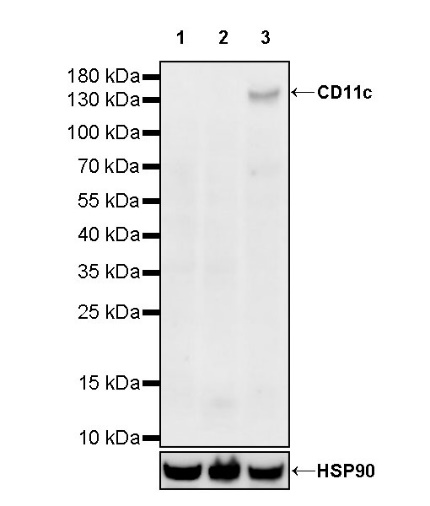
WB result of Anti-Human CD11c Mouse mAb
Primary antibody: Anti-Human CD11c Mouse mAb at 1/1000 dilution
Lane 1: Raji whole cell lysate 20 µg
Lane 2: Jurkat whole cell lysate 20 µg
Lane 3: THP-1 whole cell lysate 20 µg
Negative control: Raji whole cell lysate; Jurkat whole cell lysate
Secondary antibody: Goat Anti-mouse IgG, (H+L), HRP conjugated at 1/10000 dilution
Predicted MW: 145 kDa
Observed MW: 145 kDa (This blot was developed with high sensitivity substrate)
Flow cytometric analysis of human PBMC (human peripheral blood mononuclear cell) labelling Human CD11c antibody at 1/2000 (0.1 μg) dilution (Right) compared with a Mouse monoclonal IgG isotype control (Left). Goat Anti - Mouse IgG Alexa Fluor® 488 was used as the secondary antibody. Then cells were stained with CD14 - Alexa Fluor® 647 separately. Gated on total viable cells.
ICC shows positive staining in THP-1 cells (top panel) and negative staining in Jurkat cells (below panel). Anti- CD11c antibody was used at 1/500 dilution (Green) and incubated overnight at 4°C. Goat polyclonal Antibody to Mouse IgG - H&L (Alexa Fluor® 488) was used as secondary antibody at 1/1000 dilution. The cells were fixed with 4% PFA and permeabilized with 0.1% PBS-Triton X-100. Nuclei were counterstained with DAPI (Blue). Counterstain with tubulin (Red).
 您现在的位置: