
12 months from date of receipt / reconstitution, -20 °C as supplied
| 应用 | 稀释度 |
|---|---|
| WB | 1:1000 |
| ICC | 1:500 |
| IP | 1:50 |
| IHC-P | 1:500 |
| ICFCM | 1:500 |
HLA-DP alpha1 belongs to the HLA class II alpha chain paralogues. The class II molecule is a heterodimer consisting of an alpha (DPA) and a beta chain (DPB), both anchored in the membrane. It plays a central role in the immune system by presenting peptides derived from extracellular proteins. Class II molecules are expressed in antigen-presenting cells.
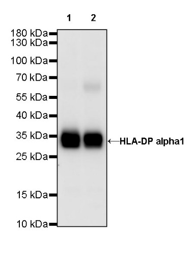
WB result of HLA-DP alpha1 Rabbit mAb
Primary antibody: HLA-DP alpha1 Rabbit mAb at 1/1000 dilution
Lane 1: Daudi whole cell lysate 20 µg
Lane 2: Ramos whole cell lysate 20 µg
Secondary antibody: Goat Anti-Rabbit IgG, (H+L), HRP conjugated at 1/10000 dilution
Predicted MW: 29 kDa
Observed MW: 33 kDa
Flow cytometric analysis of 4% PFA fixed 90% methanol permeabilized Ramos (Human Burkitt's lymphoma B lymphocyte) cells labeling HLA-DP alpha1 at 1/500 dilution (0.1 μg) / (red) compared with a rabbit monoclonal IgG isotype control (black) and an unlabeled control (cells without incubation with primary antibody and secondary antibody) (Blue). Goat Anti - Rabbit IgG Alexa Fluor® 488 was used as the secondary antibody.
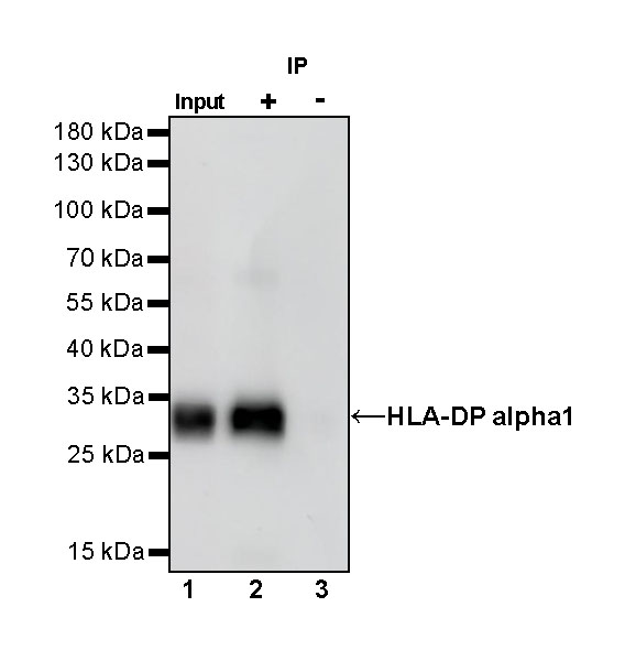
HLA-DP alpha1 Rabbit mAb at 1/50 dilution (1 µg) immunoprecipitating HLA-DP alpha1 in 0.4 mg Ramos whole cell lysate.
Western blot was performed on the immunoprecipitate using HLA-DP alpha1 Rabbit mAb at 1/1000 dilution.
Secondary antibody (HRP) for IP was used at 1/400 dilution.
Lane 1: Ramos whole cell lysate 20 µg (Input)
Lane 2: HLA-DP alpha1 Rabbit mAb IP in Ramos whole cell lysate
Lane 3: Rabbit monoclonal IgG IP in Ramos whole cell lysate
Predicted MW: 29 kDa
Observed MW: 33 kDa
IHC shows positive staining in paraffin-embedded human colon. Anti-HLA-DP alpha1 antibody was used at 1/500 dilution, followed by a HRP Polymer for Mouse & Rabbit IgG (ready to use). Counterstained with hematoxylin. Heat mediated antigen retrieval with Tris/EDTA buffer pH9.0 was performed before commencing with IHC staining protocol.
IHC shows positive staining in paraffin-embedded human tonsil. Anti-HLA-DP alpha1 antibody was used at 1/500 dilution, followed by a HRP Polymer for Mouse & Rabbit IgG (ready to use). Counterstained with hematoxylin. Heat mediated antigen retrieval with Tris/EDTA buffer pH9.0 was performed before commencing with IHC staining protocol.
IHC shows positive staining in paraffin-embedded human prostate. Anti-HLA-DP alpha1 antibody was used at 1/500 dilution, followed by a HRP Polymer for Mouse & Rabbit IgG (ready to use). Counterstained with hematoxylin. Heat mediated antigen retrieval with Tris/EDTA buffer pH9.0 was performed before commencing with IHC staining protocol.
IHC shows positive staining in paraffin-embedded human colon cancer. Anti-HLA-DP alpha1 antibody was used at 1/500 dilution, followed by a HRP Polymer for Mouse & Rabbit IgG (ready to use). Counterstained with hematoxylin. Heat mediated antigen retrieval with Tris/EDTA buffer pH9.0 was performed before commencing with IHC staining protocol.
IHC shows positive staining in paraffin-embedded human gastric cancer. Anti-HLA-DP alpha1 antibody was used at 1/500 dilution, followed by a HRP Polymer for Mouse & Rabbit IgG (ready to use). Counterstained with hematoxylin. Heat mediated antigen retrieval with Tris/EDTA buffer pH9.0 was performed before commencing with IHC staining protocol.
ICC shows positive staining in Ramos cells. Anti-HLA-DP alpha1 antibody was used at 1/500 dilution (Green) and incubated overnight at 4°C. Goat polyclonal Antibody to Rabbit IgG - H&L (Alexa Fluor® 488) was used as secondary antibody at 1/1000 dilution. The cells were fixed with 100% ice-cold methanol and permeabilized with 0.1% PBS-Triton X-100. Nuclei were counterstained with DAPI (Blue). Counterstain with tubulin (red).
 您现在的位置: