
12 months from date of receipt / reconstitution, -20 °C as supplied
| 应用 | 稀释度 |
|---|---|
| WB | 1:1000 |
| IP | 1:50 |
| IHC | 1:2000 |
| ICC | 1:500 |
| ICFCM | 1:50 |
Proliferating cell nuclear antigen (PCNA) is a type of DNA clamp that acts as a DNA polymerase in eukaryotic cells δ. The processing factor of is crucial for replication. PCNA is a homologous trimer that achieves its processability by surrounding DNA, where it serves as a scaffold for recruiting proteins involved in DNA replication, DNA repair, chromatin remodeling, and epigenetics. It is expressed in most tumors as a proliferative antigen.
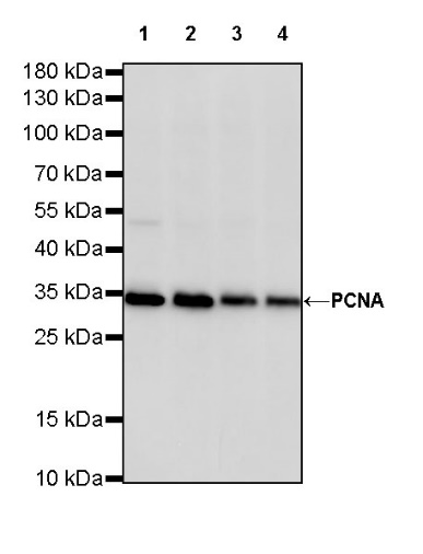
WB result of PCNA Rabbit mAb
Primary antibody: PCNA Rabbit mAb at 1/1000 dilution
Lane 1: HeLa whole cell lysate 20 µg
Lane 2: HepG2 whole cell lysate 20 µg
Lane 3: A431 whole cell lysate 20 µg
Lane 4: MCF7 whole cell lysate 20 µg
Secondary antibody: Goat Anti-Rabbit IgG, (H+L), HRP conjugated at 1/10000 dilution
Predicted MW: 29 kDa
Observed MW: 33 kDa
WB result of PCNA Rabbit mAb
Primary antibody: PCNA Rabbit mAb at 1/1000 dilution
Lane 1: mouse spleen lysate 20 µg
Lane 2: mouse testis lysate 20 µg
Lane 3: NIH/3T3 whole cell lysate 20 µg
Lane 4: C2C12 whole cell lysate 20 µg
Secondary antibody: Goat Anti-Rabbit IgG, (H+L), HRP conjugated at 1/10000 dilution
Predicted MW: 29 kDa
Observed MW: 33 kDa
WB result of PCNA Rabbit mAb
Primary antibody: PCNA Rabbit mAb at 1/1000 dilution
Lane 1: PC-12 whole cell lysate 20 µg
Lane 2: C6 whole cell lysate 20 µg
Secondary antibody: Goat Anti-Rabbit IgG, (H+L), HRP conjugated at 1/10000 dilution
Predicted MW: 29 kDa
Observed MW: 33 kDa
Flow cytometric analysis of 4% PFA fixed 90% methanol permeabilized HeLa (Human cervix adenocarcinoma epithelial cell) cells labelling PCNA antibody at 1/50 dilution (1 μg) / (red) compared with a Rabbit monoclonal IgG (Black) isotype control and an unlabelled control (cells without incubation with primary antibody and secondary antibody) (Blue). Goat Anti - Rabbit IgG Alexa Fluor® 488 was used as the secondary antibody.
PCNA Rabbit mAb at 1/50 dilution (1 µg) immunoprecipitating PCNA in 0.4 mg HeLa whole cell lysate.
Western blot was performed on the immunoprecipitate using PCNA Rabbit mAb at 1/1000 dilution.
Secondary antibody (HRP) for IP was used at 1/400 dilution.
Lane 1: HeLa whole cell lysate 20 µg (Input)
Lane 2: PCNA Rabbit mAb IP in HeLa whole cell lysate
Lane 3: Rabbit monoclonal IgG IP in HeLa whole cell lysate
Predicted MW: 29 kDa
Observed MW: 33 kDa
IHC shows positive staining in paraffin-embedded human colon. Anti-PCAN antibody was used at 1/2000 dilution, followed by a HRP Polymer for Mouse & Rabbit IgG (ready to use). Counterstained with hematoxylin. Heat mediated antigen retrieval with Tris/EDTA buffer pH9.0 was performed before commencing with IHC staining protocol.
IHC shows positive staining in paraffin-embedded human testis. Anti-PCAN antibody was used at 1/2000 dilution, followed by a HRP Polymer for Mouse & Rabbit IgG (ready to use). Counterstained with hematoxylin. Heat mediated antigen retrieval with Tris/EDTA buffer pH9.0 was performed before commencing with IHC staining protocol.
IHC shows positive staining in paraffin-embedded human hepatocellular carcinoma. Anti-PCAN antibody was used at 1/2000 dilution, followed by a HRP Polymer for Mouse & Rabbit IgG (ready to use). Counterstained with hematoxylin. Heat mediated antigen retrieval with Tris/EDTA buffer pH9.0 was performed before commencing with IHC staining protocol.
IHC shows positive staining in paraffin-embedded human bladder cancer. Anti-PCAN antibody was used at 1/2000 dilution, followed by a HRP Polymer for Mouse & Rabbit IgG (ready to use). Counterstained with hematoxylin. Heat mediated antigen retrieval with Tris/EDTA buffer pH9.0 was performed before commencing with IHC staining protocol.
IHC shows positive staining in paraffin-embedded human thyroid cancer. Anti-PCAN antibody was used at 1/2000 dilution, followed by a HRP Polymer for Mouse & Rabbit IgG (ready to use). Counterstained with hematoxylin. Heat mediated antigen retrieval with Tris/EDTA buffer pH9.0 was performed before commencing with IHC staining protocol.
IHC shows positive staining in paraffin-embedded mouse colon. Anti-PCAN antibody was used at 1/2000 dilution, followed by a HRP Polymer for Mouse & Rabbit IgG (ready to use). Counterstained with hematoxylin. Heat mediated antigen retrieval with Tris/EDTA buffer pH9.0 was performed before commencing with IHC staining protocol.
IHC shows positive staining in paraffin-embedded rat testis. Anti-PCAN antibody was used at 1/2000 dilution, followed by a HRP Polymer for Mouse & Rabbit IgG (ready to use). Counterstained with hematoxylin. Heat mediated antigen retrieval with Tris/EDTA buffer pH9.0 was performed before commencing with IHC staining protocol.
ICC shows positive staining in HeLa cells. Anti-PCNA antibody was used at 1/500 dilution (Green) and incubated overnight at 4°C. Goat polyclonal Antibody to Rabbit IgG - H&L (Alexa Fluor® 488) was used as secondary antibody at 1/1000 dilution. The cells were fixed with 4% PFA and permeabilized with 0.1% PBS-Triton X-100. Nuclei were counterstained with DAPI (Blue). Counterstain with tubulin (red).
 您现在的位置: