
12 months from date of receipt / reconstitution, -20 °C as supplied
| 应用 | 稀释度 |
|---|---|
| WB | 1:1000 |
| ICC | 1:500 |
| IP | 1:50 |
Terminal deoxynucleotidyl transferase (TdT), also known as DNA nucleotidylexotransferase (DNTT) or terminal transferase, is a specialized DNA polymerase expressed in immature, pre-B, pre-T lymphoid cells, and acute lymphoblastic leukemia/lymphoma cells. TdT adds N-nucleotides to the V, D, and J exons of the TCR and BCR genes during antibody gene recombination, enabling the phenomenon of junctional diversity. The diversity introduced by TdT has played an important role in the evolution of the vertebrate immune system, significantly increasing the variety of antigen receptors that a cell is equipped with to fight pathogens.
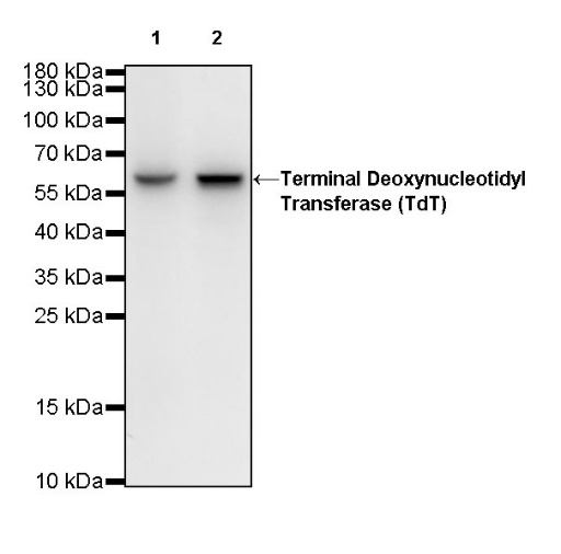
WB result of Terminal Deoxynucleotidyl Transferase (TdT) Rabbit mAb
Primary antibody: Terminal Deoxynucleotidyl Transferase (TdT) Rabbit mAb at 1/1000 dilution
Lane 1: Jurkat whole cell lysate 20 µg
Lane 2: MOLT-4 whole cell lysate 20 µg
Secondary antibody: Goat Anti-Rabbit IgG, (H+L), HRP conjugated at 1/10000 dilution
Predicted MW: 58 kDa
Observed MW: 58 kDa
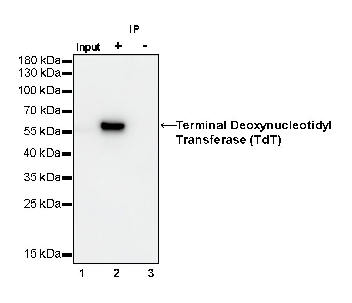
Terminal Deoxynucleotidyl Transferase (TdT) Rabbit mAb at 1/50 dilution (1 µg) immunoprecipitating Terminal Deoxynucleotidyl Transferase (TdT) in 0.4 mg Jurkat whole cell lysate.
Western blot was performed on the immunoprecipitate using Terminal Deoxynucleotidyl Transferase (TdT) Rabbit mAb at 1/1000 dilution.
Secondary antibody (HRP) for IP was used at 1/400 dilution.
Lane 1: Jurkat whole cell lysate 20 µg (Input)
Lane 2: Terminal Deoxynucleotidyl Transferase (TdT) Rabbit mAb IP in Jurkat whole cell lysate
Lane 3: Rabbit monoclonal IgG IP in Jurkat whole cell lysate
Predicted MW: 58 kDa
Observed MW: 58 kDa
(This blot was developed with high sensitivity substrate)
ICC shows positive staining in Molt-4 cells. Anti-Terminal Deoxynucleotidyl Transferase (TDT) antibody was used at 1/500 dilution (Green) and incubated overnight at 4°C. Goat polyclonal Antibody to Rabbit IgG - H&L (Alexa Fluor® 488) was used as secondary antibody at 1/1000 dilution. The cells were fixed with 4% PFA and permeabilized with 0.1% PBS-Triton X-100. Nuclei were counterstained with DAPI (Blue). Counterstain with tubulin (red).
 您现在的位置: