
12 months from date of receipt / reconstitution, -20 °C as supplied
| 应用 | 稀释度 |
|---|---|
| WB | 1:1000 |
| IP | 1:50 |
| IHC | 1:500 |
| ICC | 1:100 |
Trefoil factor 1 is a protein that in humans is encoded by the TFF1 gene. Members of the trefoil family are characterized by having at least one copy of the trefoil motif, a 40-amino acid domain that contains three conserved disulfides. They are stable secretory proteins expressed in gastrointestinal mucosa. TFF1 expression is frequently lost in gastric carcinoma, probably through mechanism of DNA methylation, and it is therefore considered as a tumor suppressor gene.
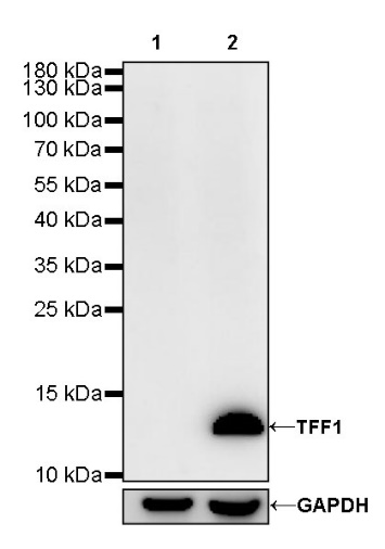
WB result of TFF1 Rabbit mAb
Primary antibody: TFF1 Rabbit mAb at 1/1000 dilution
Lane 1: MDA-MB-231 whole cell lysate 20 µg
Lane 2: MCF7 whole cell lysate 20 µg
Negative control: MDA-MB-231 whole cell lysate
Secondary antibody: Goat Anti-Rabbit IgG, (H+L), HRP conjugated at 1/10000 dilution
Predicted MW: 9 kDa
Observed MW: 13 kDa
TFF1 Rabbit mAb at 1/50 dilution (1 µg) immunoprecipitating TFF1 in 0.4 mg MCF-7 whole cell lysate.
Western blot was performed on the immunoprecipitate using TFF1 Rabbit mAb at 1/1000 dilution.
Secondary antibody (HRP) for IP was used at 1/400 dilution.
Lane 1: MCF-7 whole cell lysate 20 µg (Input)
Lane 2: TFF1 Rabbit mAb IP in MCF-7 whole cell lysate
Lane 3: Rabbit monoclonal IgG IP in MCF-7 whole cell lysate
Predicted MW: 9 kDa
Observed MW: 13 kDa
IHC shows positive staining in paraffin-embedded human stomach. Anti-TFF1 antibody was used at 1/500 dilution, followed by a HRP Polymer for Mouse & Rabbit IgG (ready to use). Counterstained with hematoxylin. Heat mediated antigen retrieval with Tris/EDTA buffer pH9.0 was performed before commencing with IHC staining protocol.
Negative control: IHC shows negative staining in paraffin-embedded human spleen. Anti-TFF1 antibody was used at 1/500 dilution, followed by a HRP Polymer for Mouse & Rabbit IgG (ready to use). Counterstained with hematoxylin. Heat mediated antigen retrieval with Tris/EDTA buffer pH9.0 was performed before commencing with IHC staining protocol.
IHC shows positive staining in paraffin-embedded human gastric cancer. Anti-TFF1 antibody was used at 1/500 dilution, followed by a HRP Polymer for Mouse & Rabbit IgG (ready to use). Counterstained with hematoxylin. Heat mediated antigen retrieval with Tris/EDTA buffer pH9.0 was performed before commencing with IHC staining protocol.
IHC shows positive staining in paraffin-embedded human pancreatic cancer. Anti-TFF1 antibody was used at 1/500 dilution, followed by a HRP Polymer for Mouse & Rabbit IgG (ready to use). Counterstained with hematoxylin. Heat mediated antigen retrieval with Tris/EDTA buffer pH9.0 was performed before commencing with IHC staining protocol.
IHC shows positive staining in paraffin-embedded human transitional cell carcinoma. Anti-TFF1 antibody was used at 1/500 dilution, followed by a HRP Polymer for Mouse & Rabbit IgG (ready to use). Counterstained with hematoxylin. Heat mediated antigen retrieval with Tris/EDTA buffer pH9.0 was performed before commencing with IHC staining protocol.
ICC shows positive staining in MCF7 cells. Anti-TFF1 antibody was used at 1/100 dilution (Green) and incubated overnight at 4°C. Goat polyclonal Antibody to Rabbit IgG - H&L (Alexa Fluor® 488) was used as secondary antibody at 1/1000 dilution. The cells were fixed with 4% PFA and permeabilized with 0.1% PBS-Triton X-100. Nuclei were counterstained with DAPI (Blue). Counterstain with tubulin (red).
 您现在的位置: